 Movie
Movie Controller

[English] 日本語
 Yorodumi
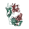
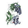
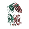
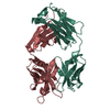
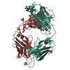
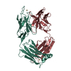
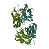
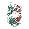
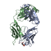
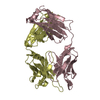
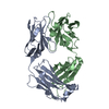
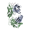
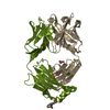
- PDB-1s5i: Fab (LNKB-2) of monoclonal antibody to Human Interleukin-2, cryst... -
+ Open data

- Basic information
| Entry | Database: PDB / ID: 1s5i | ||||||
|---|---|---|---|---|---|---|---|
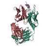
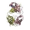
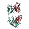
| Title | Fab (LNKB-2) of monoclonal antibody to Human Interleukin-2, crystal structure | ||||||
 Components Components | (Fab-fragment of monoclonal antibody) x 2 | ||||||
 Keywords Keywords | IMMUNE SYSTEM / monoclonal antibody / antigen-binding fragment / interleukin-2 / x-ray analysis | ||||||
| Function / homology |  Function and homology information Function and homology informationInitial triggering of complement / Classical antibody-mediated complement activation / FCGR activation / Role of phospholipids in phagocytosis / Regulation of Complement cascade / phagocytosis, recognition / humoral immune response mediated by circulating immunoglobulin / Regulation of actin dynamics for phagocytic cup formation / positive regulation of type IIa hypersensitivity / positive regulation of type I hypersensitivity ...Initial triggering of complement / Classical antibody-mediated complement activation / FCGR activation / Role of phospholipids in phagocytosis / Regulation of Complement cascade / phagocytosis, recognition / humoral immune response mediated by circulating immunoglobulin / Regulation of actin dynamics for phagocytic cup formation / positive regulation of type IIa hypersensitivity / positive regulation of type I hypersensitivity / antibody-dependent cellular cytotoxicity / Fc-gamma receptor I complex binding / immunoglobulin complex, circulating / phagocytosis, engulfment / immunoglobulin receptor binding / IgG immunoglobulin complex / immunoglobulin mediated immune response / complement activation, classical pathway / antigen binding / positive regulation of phagocytosis / B cell differentiation / positive regulation of immune response / antibacterial humoral response / defense response to bacterium / external side of plasma membrane / : / cytoplasm Similarity search - Function | ||||||
| Biological species |  | ||||||
| Method |  X-RAY DIFFRACTION / MOLECULAR REPLACEMENT / Resolution: 2.7 Å X-RAY DIFFRACTION / MOLECULAR REPLACEMENT / Resolution: 2.7 Å | ||||||
 Authors Authors | Pletnev, V.Z. / Goryacheva, E.A. / Tsygannik, I.N. / Nesmeyanov, V.A. / Pletnev, S.V. / Pangborn, W. / Duax, W. | ||||||
 Citation Citation | Journal: Bioorg. Khim. Title: [A new crystal form of the Fab fragment of a monoclonal antibody to human interleukin-2: the three-dimensional structure at 2.7 A resolution]. Authors: Pletnev, V.Z. / Goriacheva, E.A. / Tsygannik, I.N. / Nesmeianov, V.A. / Pletnev, S.V. / Pangborn, W. / Daux, W. #1: Journal: Bioorg.Khim. / Year: 2000Title: Spatial structure of a Fab-fragment of a monoclonal antibody to human interleukin-2 in two crystalline forms at a resolution of 2.2 and 2.9 angstroms Authors: Fokin, A.V. / Afonin, P.V. / Mikhailova, I.Y. / Tsygannik, I.N. / Mareeva, T.Y. / Nesmeyanov, V.A. / Pangborn, W. / Lee, N. / Duax, W. / Siszak, E. / Pletnev, V.Z. #2: Journal: Protein Sci. / Year: 2001Title: Crystal structure of an anti-interleukin-2 monoclonal antibody Fab complexed with an antigenic nonapeptide Authors: Afonin, P.V. / Fokin, A.V. / Tsygannik, I.N. / Mikhailova, I.Y. / Onoprienko, L.V. / Michaleva, I.I. / Ivanov, V.T. / Mareeva, T.Y. / Nesmeyanov, V.A. / Li, N. / Pangborn, W. / Duax, W. / Pletnev, V.Z. | ||||||
| History |
| ||||||
| Remark 999 | SEQUENCE The sequence of this protein was not deposited into any sequence database. |
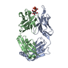
- Structure visualization
| Structure viewer | Molecule: MolmilJmol/JSmol |
|---|
- Downloads & links
-Download
| PDBx/mmCIF format | 1s5i.cif.gz | 97.3 KB | Display | PDBx/mmCIF format |
|---|---|---|---|---|
| PDB format | pdb1s5i.ent.gz | 74.3 KB | Display | PDB format |
| PDBx/mmJSON format | 1s5i.json.gz | Tree view | PDBx/mmJSON format | |
| Others |  Other downloads Other downloads |
-Validation report
| Arichive directory | https://data.pdbj.org/pub/pdb/validation_reports/s5/1s5iftp://data.pdbj.org/pub/pdb/validation_reports/s5/1s5i | HTTPS FTP |
|---|
-Related structure data
| Related structure data | 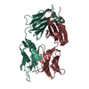 1f8tS S: Starting model for refinement |
|---|---|
| Similar structure data |


-Links
 PDBj
PDBj


- Assembly
| Deposited unit | 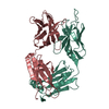
| ||||||||
|---|---|---|---|---|---|---|---|---|---|
| 1 |
| ||||||||
| Unit cell |
|
-Components
| #1: Antibody | Mass: 24203.908 Da / Num. of mol.: 1 / Source method: isolated from a natural source / Source: (natural) |
|---|---|
| #2: Antibody | Mass: 23869.600 Da / Num. of mol.: 1 / Source method: isolated from a natural source / Source: (natural) |
| #3: Water | ChemComp-HOH /  Mass: 18.015 Da / Num. of mol.: 66 / Source method: isolated from a natural source / Formula: H2O Mass: 18.015 Da / Num. of mol.: 66 / Source method: isolated from a natural source / Formula: H2O |
| Has protein modification | Y |
-Experimental details
-Experiment
| Experiment | Method: X-RAY DIFFRACTION / Number of used crystals: 1 |
|---|
- Sample preparation
| Crystal | Density Matthews: 2.87 Å3/Da / Density % sol: 56.76 % |
|---|---|
| Crystal grow | Temperature: 300 K / Method: vapor diffusion, hanging drop / pH: 6.5 Details: PEG-8000, ZnAc, NaN3, Na(CH3)2AsO4, pH 6.5, VAPOR DIFFUSION, HANGING DROP, temperature 300K |
-Data collection
| Diffraction | Mean temperature: 300 K |
|---|---|
| Diffraction source | Source: ROTATING ANODE / Type: RIGAKU RU200 / Wavelength: 1.5418 Å |
| Detector | Type: RIGAKU RAXIS IIC / Detector: IMAGE PLATE / Date: Jan 1, 2001 |
| Radiation | Monochromator: GRAPHITE / Protocol: SINGLE WAVELENGTH / Monochromatic (M) / Laue (L): M / Scattering type: x-ray |
| Radiation wavelength | Wavelength: 1.5418 Å / Relative weight: 1 |
| Reflection | Resolution: 2.7→41.5 Å / Num. all: 14370 / Num. obs: 14370 / % possible obs: 100 % / Observed criterion σ(F): 0 / Redundancy: 3.5 % / Rmerge(I) obs: 0.072 / Net I/σ(I): 8.1 |
| Reflection shell | Resolution: 2.7→2.76 Å / Redundancy: 3.3 % / Rmerge(I) obs: 0.32 / Num. unique all: 879 / % possible all: 88.3 |
- Processing
| Software |
| |||||||||||||||||||||||||
|---|---|---|---|---|---|---|---|---|---|---|---|---|---|---|---|---|---|---|---|---|---|---|---|---|---|---|
| Refinement | Method to determine structure: MOLECULAR REPLACEMENT Starting model: PDB ENTRY 1F8T Resolution: 2.7→41.5 Å / Isotropic thermal model: isotropic / Cross valid method: THROUGHOUT / σ(F): 0 / Stereochemistry target values: Engh & Huber Details: Cross validation maximum likelihood simulated annealing refinement (CNS package)
| |||||||||||||||||||||||||
| Displacement parameters | Biso mean: 38.8 Å2
| |||||||||||||||||||||||||
| Refine analyze | Luzzati coordinate error obs: 0.43 Å / Luzzati sigma a obs: 0.55 Å | |||||||||||||||||||||||||
| Refinement step | Cycle: LAST / Resolution: 2.7→41.5 Å
| |||||||||||||||||||||||||
| Refine LS restraints |
| |||||||||||||||||||||||||
| Xplor file |
|
