 Movie
Movie Controller


+ Open data

- Basic information
| Entry | Database: PDB / ID: 2a77 | ||||||
|---|---|---|---|---|---|---|---|
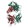
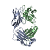
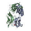
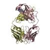
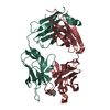
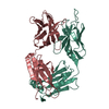
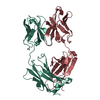
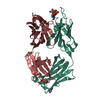
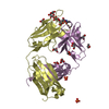
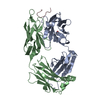
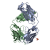
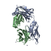
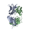
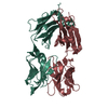
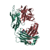
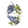
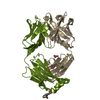
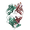
| Title | Anti-Cocaine Antibody 7.5.21, Crystal Form II | ||||||
 Components Components |
| ||||||
 Keywords Keywords | IMMUNE SYSTEM | ||||||
| Function / homology | Immunoglobulins / Immunoglobulin-like / Sandwich / Mainly Beta Function and homology information Function and homology information | ||||||
| Biological species |  | ||||||
| Method |  X-RAY DIFFRACTION / SYNCHROTRON / MOLECULAR REPLACEMENT / Resolution: 1.8 Å X-RAY DIFFRACTION / SYNCHROTRON / MOLECULAR REPLACEMENT / Resolution: 1.8 Å | ||||||
 Authors Authors | Pozharski, E. / Hewagama, A. / Shanafelt, A. / Ringe, D. / Petsko, G.A. | ||||||
 Citation Citation | Journal: To be Published Title: Flexibility of Packing: Four Crystal Forms of an Anti-Cocaine Antibody 7.5.21 Authors: Pozharski, E. / Hewagama, A. / Shanafelt, A. / Ringe, D. / Petsko, G.A. | ||||||
| History |
|
- Structure visualization
| Structure viewer | Molecule: MolmilJmol/JSmol |
|---|
- Downloads & links
-Download
| PDBx/mmCIF format | 2a77.cif.gz | 107.7 KB | Display | PDBx/mmCIF format |
|---|---|---|---|---|
| PDB format | pdb2a77.ent.gz | 81.6 KB | Display | PDB format |
| PDBx/mmJSON format | 2a77.json.gz | Tree view | PDBx/mmJSON format | |
| Others |  Other downloads Other downloads |
-Validation report
| Arichive directory | https://data.pdbj.org/pub/pdb/validation_reports/a7/2a77ftp://data.pdbj.org/pub/pdb/validation_reports/a7/2a77 | HTTPS FTP |
|---|
-Related structure data
| Related structure data |  2ai0C 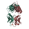 2a1wS S: Starting model for refinement C: citing same article ( |
|---|---|
| Similar structure data |


-Links
 PDBj
PDBj

- Assembly
| Deposited unit | 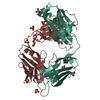
| ||||||||
|---|---|---|---|---|---|---|---|---|---|
| 1 |
| ||||||||
| 2 | 
| ||||||||
| Unit cell |
|
-Components
| #1: Antibody | Mass: 23893.607 Da / Num. of mol.: 1 / Source method: isolated from a natural source / Source: (natural) | ||||||
|---|---|---|---|---|---|---|---|
| #2: Antibody | Mass: 23868.779 Da / Num. of mol.: 1 / Source method: isolated from a natural source / Source: (natural) | ||||||
| #3: Chemical | ChemComp-SO4 /   Mass: 96.063 Da / Num. of mol.: 6 / Source method: obtained synthetically / Formula: SO4 Mass: 96.063 Da / Num. of mol.: 6 / Source method: obtained synthetically / Formula: SO4#4: Chemical | ChemComp-GOL /   Mass: 92.094 Da / Num. of mol.: 7 / Source method: obtained synthetically / Formula: C3H8O3 Mass: 92.094 Da / Num. of mol.: 7 / Source method: obtained synthetically / Formula: C3H8O3#5: Water | ChemComp-HOH / |  Mass: 18.015 Da / Num. of mol.: 318 / Source method: isolated from a natural source / Formula: H2O Mass: 18.015 Da / Num. of mol.: 318 / Source method: isolated from a natural source / Formula: H2OHas protein modification | Y | |
-Experimental details
-Experiment
| Experiment | Method: X-RAY DIFFRACTION |
|---|
- Sample preparation
| Crystal | Density Matthews: 2.7 Å3/Da / Density % sol: 54.7 % |
|---|---|
| Crystal grow | Temperature: 298 K / Method: vapor diffusion, hanging drop / pH: 7 Details: 16% PEG8000, 0.3M ammonium sulfate, pH 7.0, VAPOR DIFFUSION, HANGING DROP, temperature 298K |
-Data collection
| Diffraction | Mean temperature: 105 K |
|---|---|
| Diffraction source | Source: SYNCHROTRON / Site: SSRL  / Beamline: BL11-1 / Wavelength: 0.89996 Å / Beamline: BL11-1 / Wavelength: 0.89996 Å |
| Detector | Type: ADSC QUANTUM 4 / Detector: CCD / Date: Jul 4, 2004 |
| Radiation | Protocol: SINGLE WAVELENGTH / Monochromatic (M) / Laue (L): M / Scattering type: x-ray |
| Radiation wavelength | Wavelength: 0.89996 Å / Relative weight: 1 |
| Reflection | Resolution: 1.8→50 Å / Num. all: 46600 / Num. obs: 46600 / % possible obs: 97 % / Observed criterion σ(F): 0 / Redundancy: 4 % / Biso Wilson estimate: 33.3 Å2 / Rmerge(I) obs: 0.044 / Χ2: 1.124 / Net I/σ(I): 33 |
| Reflection shell | Resolution: 1.8→1.86 Å / % possible obs: 87.2 % / Redundancy: 3.4 % / Mean I/σ(I) obs: 1.7 / Num. measured obs: 4182 / Num. unique all: 4182 / Χ2: 0.971 / % possible all: 87.2 |
- Processing
| Software |
| ||||||||||||||||||||||||||||||||||||||||||||||||||||||||||||||||||||||||||||||||||||||||||
|---|---|---|---|---|---|---|---|---|---|---|---|---|---|---|---|---|---|---|---|---|---|---|---|---|---|---|---|---|---|---|---|---|---|---|---|---|---|---|---|---|---|---|---|---|---|---|---|---|---|---|---|---|---|---|---|---|---|---|---|---|---|---|---|---|---|---|---|---|---|---|---|---|---|---|---|---|---|---|---|---|---|---|---|---|---|---|---|---|---|---|---|
| Refinement | Method to determine structure: MOLECULAR REPLACEMENT Starting model: PDB ENTRY 2A1W Resolution: 1.8→33.3 Å / Cor.coef. Fo:Fc: 0.948 / Cor.coef. Fo:Fc free: 0.926 / SU B: 8.041 / SU ML: 0.123 / TLS residual ADP flag: LIKELY RESIDUAL / Cross valid method: THROUGHOUT / σ(F): 0 / ESU R: 0.144 / ESU R Free: 0.142 / Stereochemistry target values: MAXIMUM LIKELIHOOD
| ||||||||||||||||||||||||||||||||||||||||||||||||||||||||||||||||||||||||||||||||||||||||||
| Solvent computation | Ion probe radii: 0.8 Å / Shrinkage radii: 0.8 Å / VDW probe radii: 1.2 Å / Solvent model: MASK | ||||||||||||||||||||||||||||||||||||||||||||||||||||||||||||||||||||||||||||||||||||||||||
| Displacement parameters | Biso mean: 40.947 Å2
| ||||||||||||||||||||||||||||||||||||||||||||||||||||||||||||||||||||||||||||||||||||||||||
| Refinement step | Cycle: LAST / Resolution: 1.8→33.3 Å
| ||||||||||||||||||||||||||||||||||||||||||||||||||||||||||||||||||||||||||||||||||||||||||
| Refine LS restraints |
| ||||||||||||||||||||||||||||||||||||||||||||||||||||||||||||||||||||||||||||||||||||||||||
| LS refinement shell | Resolution: 1.799→1.846 Å / Total num. of bins used: 20
| ||||||||||||||||||||||||||||||||||||||||||||||||||||||||||||||||||||||||||||||||||||||||||
| Refinement TLS params. | Method: refined / Refine-ID: X-RAY DIFFRACTION
| ||||||||||||||||||||||||||||||||||||||||||||||||||||||||||||||||||||||||||||||||||||||||||
| Refinement TLS group | Refine-ID: X-RAY DIFFRACTION / Selection: ALL
|
