 Movie
Movie Controller


+ Open data

- Basic information
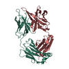
| Entry | Database: PDB / ID: 1f8t | ||||||
|---|---|---|---|---|---|---|---|
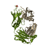
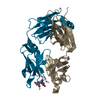
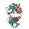
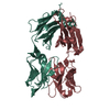
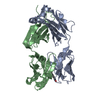
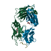
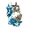
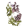
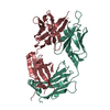
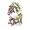
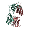
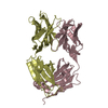
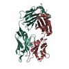
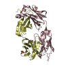
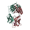
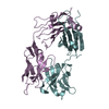
| Title | FAB (LNKB-2) OF MONOCLONAL ANTIBODY, CRYSTAL STRUCTURE | ||||||
 Components Components |
| ||||||
 Keywords Keywords | IMMUNE SYSTEM / monoclonal antibody / antigen-binding fragment / interleukin-2 / x-ray analysis / crystal | ||||||
| Function / homology | Immunoglobulins / Immunoglobulin-like / Sandwich / Mainly Beta Function and homology information Function and homology information | ||||||
| Biological species |  | ||||||
| Method |  X-RAY DIFFRACTION / Resolution: 2.2 Å X-RAY DIFFRACTION / Resolution: 2.2 Å | ||||||
 Authors Authors | Fokin, A.V. / Afonin, P.V. / Mikhailova, I.I. / Tsygannik, I.N. / Mareeva, T.I. | ||||||
 Citation Citation | Journal: Bioorg.Khim. / Year: 2000 Title: Spatial structure of a Fab-fragment of a monoclonal antibody to human interleukin-2 in two crystalline forms at a resolution of 2.2 and 2.9 angstroms Authors: Fokin, A.V. / Afonin, P.V. / Mikhailova, I.I.u. / Tsygannik, I.N. / Mareeva, T.I.u. / Nesmeianov, V.A. / Pangborn, W. / Lee, N. / Duax, W. / Siszak, E. / Pletnev, V.Z. | ||||||
| History |
|
- Structure visualization
| Structure viewer | Molecule: MolmilJmol/JSmol |
|---|
- Downloads & links
-Download
| PDBx/mmCIF format | 1f8t.cif.gz | 101.6 KB | Display | PDBx/mmCIF format |
|---|---|---|---|---|
| PDB format | pdb1f8t.ent.gz | 77.6 KB | Display | PDB format |
| PDBx/mmJSON format | 1f8t.json.gz | Tree view | PDBx/mmJSON format | |
| Others |  Other downloads Other downloads |
-Validation report
| Arichive directory | https://data.pdbj.org/pub/pdb/validation_reports/f8/1f8tftp://data.pdbj.org/pub/pdb/validation_reports/f8/1f8t | HTTPS FTP |
|---|
-Related structure data
| Similar structure data |
|---|


-Links
 PDBj
PDBj

- Assembly
| Deposited unit | 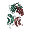
| ||||||||
|---|---|---|---|---|---|---|---|---|---|
| 1 |
| ||||||||
| Unit cell |
|
-Components
| #1: Antibody | Mass: 24203.908 Da / Num. of mol.: 1 / Source method: isolated from a natural source / Source: (natural) |
|---|---|
| #2: Antibody | Mass: 23853.600 Da / Num. of mol.: 1 / Source method: isolated from a natural source / Source: (natural) |
| #3: Water | ChemComp-HOH /  Mass: 18.015 Da / Num. of mol.: 213 / Source method: isolated from a natural source / Formula: H2O Mass: 18.015 Da / Num. of mol.: 213 / Source method: isolated from a natural source / Formula: H2O |
| Has protein modification | Y |
-Experimental details
-Experiment
| Experiment | Method: X-RAY DIFFRACTION / Number of used crystals: 1 |
|---|
- Sample preparation
| Crystal | Density Matthews: 2.35 Å3/Da / Density % sol: 47.72 % |
|---|---|
| Crystal grow | Temperature: 300 K / Method: vapor diffusion, hanging drop / pH: 5.6 Details: PEG-4000, 2-propanol, Na-citrate, pH 5.6, VAPOR DIFFUSION, HANGING DROP, temperature 300.0K |
-Data collection
| Diffraction | Mean temperature: 300 K |
|---|---|
| Diffraction source | Source: ROTATING ANODE / Type: RIGAKU RU200 / Wavelength: 1.5418 |
| Detector | Type: RIGAKU RAXIS IIC / Detector: IMAGE PLATE / Date: Feb 22, 1999 |
| Radiation | Protocol: SINGLE WAVELENGTH / Monochromatic (M) / Laue (L): M / Scattering type: x-ray |
| Radiation wavelength | Wavelength: 1.5418 Å / Relative weight: 1 |
| Reflection | Resolution: 2.2→99 Å / Num. all: 22492 / Num. obs: 21459 / % possible obs: 90.6 % / Observed criterion σ(F): 2 / Redundancy: 5.7 % / Rmerge(I) obs: 0.048 / Net I/σ(I): 24.2 |
| Reflection shell | Resolution: 2.2→2.31 Å / Redundancy: 2.8 % / Rmerge(I) obs: 0.1935 / Num. unique all: 3156 / % possible all: 94.2 |
- Processing
| Software |
| |||||||||||||||||||||||||
|---|---|---|---|---|---|---|---|---|---|---|---|---|---|---|---|---|---|---|---|---|---|---|---|---|---|---|
| Refinement | Resolution: 2.2→99 Å / σ(F): 2 / Stereochemistry target values: Engh & Huber Details: Cross-validated maximum likelihood simulated annealing refinement (CNS package)
| |||||||||||||||||||||||||
| Refinement step | Cycle: LAST / Resolution: 2.2→99 Å
| |||||||||||||||||||||||||
| Refine LS restraints |
|
