 Movie
Movie Controller

[English] 日本語
 Yorodumi
- PDB-6me1: Crystal structure of the clade B isolate B41 mutant fusion peptid... -
+ Open data

- Basic information
| Entry | Database: PDB / ID: 6me1 | ||||||
|---|---|---|---|---|---|---|---|
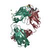
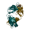
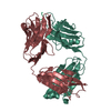
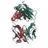
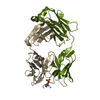
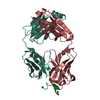
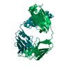
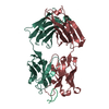
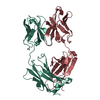
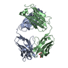
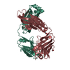
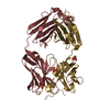
| Title | Crystal structure of the clade B isolate B41 mutant fusion peptide (residues 512-521) in complex with VRC34.01 | ||||||
 Components Components |
| ||||||
 Keywords Keywords | VIRAL PROTEIN / HIV / ENVELOPE / FUSION PEPTIDE / HIV-1 GP41 / IMMUNE SYSTEM / NEUTRALIZING ANTIBODIES / COMPLEX / VIRAL PROTEIN-IMMUNE SYSTEM COMPLEX | ||||||
| Function / homology |  Function and homology information Function and homology informationhost cell periphery / CD4 receptor binding / Dectin-2 family / symbiont-mediated perturbation of host defense response / positive regulation of plasma membrane raft polarization / positive regulation of receptor clustering / host cell endosome membrane / clathrin-dependent endocytosis of virus by host cell / viral protein processing / host cell perinuclear region of cytoplasm ...host cell periphery / CD4 receptor binding / Dectin-2 family / symbiont-mediated perturbation of host defense response / positive regulation of plasma membrane raft polarization / positive regulation of receptor clustering / host cell endosome membrane / clathrin-dependent endocytosis of virus by host cell / viral protein processing / host cell perinuclear region of cytoplasm / fusion of virus membrane with host plasma membrane / fusion of virus membrane with host endosome membrane / viral envelope / virion attachment to host cell / host cell plasma membrane / protein-containing complex binding / virion membrane / structural molecule activity / membrane Similarity search - Function | ||||||
| Biological species |  Homo sapiens (human) Homo sapiens (human)  Human immunodeficiency virus type 1 Human immunodeficiency virus type 1 | ||||||
| Method |  X-RAY DIFFRACTION / SYNCHROTRON / MOLECULAR REPLACEMENT / Resolution: 1.97 Å X-RAY DIFFRACTION / SYNCHROTRON / MOLECULAR REPLACEMENT / Resolution: 1.97 Å | ||||||
 Authors Authors | Kumar, S. / Sarkar, A. / Wilson, I.A. | ||||||
 Citation Citation | Journal: Nat Commun / Year: 2019 Title: Capturing the inherent structural dynamics of the HIV-1 envelope glycoprotein fusion peptide. Authors: Kumar, S. / Sarkar, A. / Pugach, P. / Sanders, R.W. / Moore, J.P. / Ward, A.B. / Wilson, I.A. | ||||||
| History |
|
- Structure visualization
| Structure viewer | Molecule: MolmilJmol/JSmol |
|---|
- Downloads & links
-Download
| PDBx/mmCIF format | 6me1.cif.gz | 194.9 KB | Display | PDBx/mmCIF format |
|---|---|---|---|---|
| PDB format | pdb6me1.ent.gz | 152.7 KB | Display | PDB format |
| PDBx/mmJSON format | 6me1.json.gz | Tree view | PDBx/mmJSON format | |
| Others |  Other downloads Other downloads |
-Validation report
| Arichive directory | https://data.pdbj.org/pub/pdb/validation_reports/me/6me1ftp://data.pdbj.org/pub/pdb/validation_reports/me/6me1 | HTTPS FTP |
|---|
-Related structure data
| Related structure data |  6mcoC  6mdtC  5i8cS C: citing same article ( S: Starting model for refinement |
|---|---|
| Similar structure data |


-Links
 PDBj
PDBj


- Assembly
| Deposited unit | 
| ||||||||
|---|---|---|---|---|---|---|---|---|---|
| 1 | 
| ||||||||
| 2 | 
| ||||||||
| Unit cell |
|
-Components
| #1: Antibody | Mass: 23797.615 Da / Num. of mol.: 2 Source method: isolated from a genetically manipulated source Source: (gene. exp.) Homo sapiens (human) / Production host: Homo sapiens (human)#2: Antibody | Mass: 23138.766 Da / Num. of mol.: 2 Source method: isolated from a genetically manipulated source Source: (gene. exp.) Homo sapiens (human) / Production host: Homo sapiens (human)#3: Protein/peptide | Mass: 1731.958 Da / Num. of mol.: 2 / Fragment: residues 512-521 / Source method: obtained synthetically / Source: (synth.) Human immunodeficiency virus type 1 / References: UniProt: P03375#4: Water | ChemComp-HOH / |  Mass: 18.015 Da / Num. of mol.: 572 / Source method: isolated from a natural source / Formula: H2O Mass: 18.015 Da / Num. of mol.: 572 / Source method: isolated from a natural source / Formula: H2OHas protein modification | Y | |
|---|
-Experimental details
-Experiment
| Experiment | Method: X-RAY DIFFRACTION / Number of used crystals: 1 |
|---|
- Sample preparation
| Crystal | Density Matthews: 2.75 Å3/Da / Density % sol: 55.3 % |
|---|---|
| Crystal grow | Temperature: 293.15 K / Method: vapor diffusion, sitting drop / Details: 0.2M sodium sulfate, 20%(w/v) PEG 3350 |
-Data collection
| Diffraction | Mean temperature: 100 K |
|---|---|
| Diffraction source | Source: SYNCHROTRON / Site: APS  / Beamline: 23-ID-B / Wavelength: 1.0332 Å / Beamline: 23-ID-B / Wavelength: 1.0332 Å |
| Detector | Type: DECTRIS EIGER X 16M / Detector: PIXEL / Date: Apr 6, 2018 |
| Radiation | Protocol: SINGLE WAVELENGTH / Monochromatic (M) / Laue (L): M / Scattering type: x-ray |
| Radiation wavelength | Wavelength: 1.0332 Å / Relative weight: 1 |
| Reflection | Resolution: 1.97→50 Å / Num. obs: 72903 / % possible obs: 99.3 % / Redundancy: 3.2 % / CC1/2: 0.89 / Net I/σ(I): 11.4 |
| Reflection shell | Resolution: 1.97→2.01 Å / Redundancy: 3.1 % / Mean I/σ(I) obs: 1.1 / Num. unique obs: 3639 / CC1/2: 0.64 / % possible all: 99.6 |
- Processing
| Software |
| |||||||||||||||||||||||||||||||||||||||||||||||||||||||||||||||||||||||||||||||||||||||||||||||||||||||||||||||||||||||||||||||||||||||||||||||||||||||||||||||||||||||||||||||||||||||||||||
|---|---|---|---|---|---|---|---|---|---|---|---|---|---|---|---|---|---|---|---|---|---|---|---|---|---|---|---|---|---|---|---|---|---|---|---|---|---|---|---|---|---|---|---|---|---|---|---|---|---|---|---|---|---|---|---|---|---|---|---|---|---|---|---|---|---|---|---|---|---|---|---|---|---|---|---|---|---|---|---|---|---|---|---|---|---|---|---|---|---|---|---|---|---|---|---|---|---|---|---|---|---|---|---|---|---|---|---|---|---|---|---|---|---|---|---|---|---|---|---|---|---|---|---|---|---|---|---|---|---|---|---|---|---|---|---|---|---|---|---|---|---|---|---|---|---|---|---|---|---|---|---|---|---|---|---|---|---|---|---|---|---|---|---|---|---|---|---|---|---|---|---|---|---|---|---|---|---|---|---|---|---|---|---|---|---|---|---|---|---|---|
| Refinement | Method to determine structure: MOLECULAR REPLACEMENT Starting model: 5I8C Resolution: 1.97→38.18 Å / SU ML: 0.25 / Cross valid method: FREE R-VALUE / σ(F): 1.35 / Phase error: 25.68
| |||||||||||||||||||||||||||||||||||||||||||||||||||||||||||||||||||||||||||||||||||||||||||||||||||||||||||||||||||||||||||||||||||||||||||||||||||||||||||||||||||||||||||||||||||||||||||||
| Solvent computation | Shrinkage radii: 0.9 Å / VDW probe radii: 1.11 Å | |||||||||||||||||||||||||||||||||||||||||||||||||||||||||||||||||||||||||||||||||||||||||||||||||||||||||||||||||||||||||||||||||||||||||||||||||||||||||||||||||||||||||||||||||||||||||||||
| Refinement step | Cycle: LAST / Resolution: 1.97→38.18 Å
| |||||||||||||||||||||||||||||||||||||||||||||||||||||||||||||||||||||||||||||||||||||||||||||||||||||||||||||||||||||||||||||||||||||||||||||||||||||||||||||||||||||||||||||||||||||||||||||
| Refine LS restraints |
| |||||||||||||||||||||||||||||||||||||||||||||||||||||||||||||||||||||||||||||||||||||||||||||||||||||||||||||||||||||||||||||||||||||||||||||||||||||||||||||||||||||||||||||||||||||||||||||
| LS refinement shell |
|
