 Movie
Movie Controller

[English] 日本語
 Yorodumi
- PDB-5i8c: Crystal Structure of HIV-1 Clade A BG505 Fusion Peptide (residue ... -
+ Open data

- Basic information
| Entry | Database: PDB / ID: 5i8c | ||||||
|---|---|---|---|---|---|---|---|
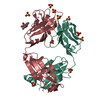
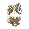
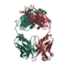
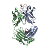
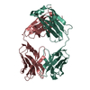
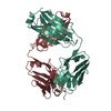
| Title | Crystal Structure of HIV-1 Clade A BG505 Fusion Peptide (residue 512-520) in Complex with Broadly Neutralizing Antibody VRC34.01 Fab | ||||||
 Components Components |
| ||||||
 Keywords Keywords | IMMUNE SYSTEM / HIV-1 / envelope / trimer / fusion peptide / antibody / neutralizing | ||||||
| Function / homology |  Function and homology information Function and homology informationsymbiont-mediated perturbation of host defense response / positive regulation of plasma membrane raft polarization / positive regulation of receptor clustering / host cell endosome membrane / clathrin-dependent endocytosis of virus by host cell / viral protein processing / fusion of virus membrane with host plasma membrane / fusion of virus membrane with host endosome membrane / viral envelope / virion attachment to host cell ...symbiont-mediated perturbation of host defense response / positive regulation of plasma membrane raft polarization / positive regulation of receptor clustering / host cell endosome membrane / clathrin-dependent endocytosis of virus by host cell / viral protein processing / fusion of virus membrane with host plasma membrane / fusion of virus membrane with host endosome membrane / viral envelope / virion attachment to host cell / host cell plasma membrane / virion membrane / structural molecule activity / membrane Similarity search - Function | ||||||
| Biological species |  Homo sapiens (human) Homo sapiens (human)  Human immunodeficiency virus 1 Human immunodeficiency virus 1 | ||||||
| Method |  X-RAY DIFFRACTION / SYNCHROTRON / MOLECULAR REPLACEMENT / Resolution: 1.54 Å X-RAY DIFFRACTION / SYNCHROTRON / MOLECULAR REPLACEMENT / Resolution: 1.54 Å | ||||||
 Authors Authors | Xu, K. / Zhou, T. / Liu, K. / Kwong, P.D. | ||||||
| Funding support |  United States, 1items United States, 1items
| ||||||
 Citation Citation | Journal: Science / Year: 2016 Title: Fusion peptide of HIV-1 as a site of vulnerability to neutralizing antibody. Authors: Rui Kong / Kai Xu / Tongqing Zhou / Priyamvada Acharya / Thomas Lemmin / Kevin Liu / Gabriel Ozorowski / Cinque Soto / Justin D Taft / Robert T Bailer / Evan M Cale / Lei Chen / Chang W Choi ...Authors: Rui Kong / Kai Xu / Tongqing Zhou / Priyamvada Acharya / Thomas Lemmin / Kevin Liu / Gabriel Ozorowski / Cinque Soto / Justin D Taft / Robert T Bailer / Evan M Cale / Lei Chen / Chang W Choi / Gwo-Yu Chuang / Nicole A Doria-Rose / Aliaksandr Druz / Ivelin S Georgiev / Jason Gorman / Jinghe Huang / M Gordon Joyce / Mark K Louder / Xiaochu Ma / Krisha McKee / Sijy O'Dell / Marie Pancera / Yongping Yang / Scott C Blanchard / Walther Mothes / Dennis R Burton / Wayne C Koff / Mark Connors / Andrew B Ward / Peter D Kwong / John R Mascola / Abstract: The HIV-1 fusion peptide, comprising 15 to 20 hydrophobic residues at the N terminus of the Env-gp41 subunit, is a critical component of the virus-cell entry machinery. Here, we report the ...The HIV-1 fusion peptide, comprising 15 to 20 hydrophobic residues at the N terminus of the Env-gp41 subunit, is a critical component of the virus-cell entry machinery. Here, we report the identification of a neutralizing antibody, N123-VRC34.01, which targets the fusion peptide and blocks viral entry by inhibiting conformational changes in gp120 and gp41 subunits of Env required for entry. Crystal structures of N123-VRC34.01 liganded to the fusion peptide, and to the full Env trimer, revealed an epitope consisting of the N-terminal eight residues of the gp41 fusion peptide and glycan N88 of gp120, and molecular dynamics showed that the N-terminal portion of the fusion peptide can be solvent-exposed. These results reveal the fusion peptide to be a neutralizing antibody epitope and thus a target for vaccine design. | ||||||
| History |
|
- Structure visualization
| Structure viewer | Molecule: MolmilJmol/JSmol |
|---|
- Downloads & links
-Download
| PDBx/mmCIF format | 5i8c.cif.gz | 117 KB | Display | PDBx/mmCIF format |
|---|---|---|---|---|
| PDB format | pdb5i8c.ent.gz | 86.5 KB | Display | PDB format |
| PDBx/mmJSON format | 5i8c.json.gz | Tree view | PDBx/mmJSON format | |
| Others |  Other downloads Other downloads |
-Validation report
| Arichive directory | https://data.pdbj.org/pub/pdb/validation_reports/i8/5i8cftp://data.pdbj.org/pub/pdb/validation_reports/i8/5i8c | HTTPS FTP |
|---|
-Related structure data
| Related structure data |  8125C  5i8eC 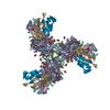 5i8hC  4jpiS C: citing same article ( S: Starting model for refinement |
|---|---|
| Similar structure data |


-Links
 PDBj
PDBj


- Assembly
| Deposited unit | 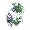
| ||||||||
|---|---|---|---|---|---|---|---|---|---|
| 1 |
| ||||||||
| Unit cell |
|
-Components
| #1: Antibody | Mass: 23797.615 Da / Num. of mol.: 1 Source method: isolated from a genetically manipulated source Source: (gene. exp.) Homo sapiens (human) / Gene: IGHG1 / Production host: Homo sapiens (human) |
|---|---|
| #2: Antibody | Mass: 23138.766 Da / Num. of mol.: 1 Source method: isolated from a genetically manipulated source Source: (gene. exp.) Homo sapiens (human) / Production host: Homo sapiens (human) |
| #3: Protein/peptide | Mass: 846.025 Da / Num. of mol.: 1 / Source method: obtained synthetically / Source: (synth.) Human immunodeficiency virus 1 / References: UniProt: Q2N0S7*PLUS |
| #4: Water | ChemComp-HOH /  Mass: 18.015 Da / Num. of mol.: 759 / Source method: isolated from a natural source / Formula: H2O Mass: 18.015 Da / Num. of mol.: 759 / Source method: isolated from a natural source / Formula: H2O |
| Has protein modification | Y |
-Experimental details
-Experiment
| Experiment | Method: X-RAY DIFFRACTION / Number of used crystals: 1 |
|---|
- Sample preparation
| Crystal | Density Matthews: 2.11 Å3/Da / Density % sol: 41.59 % |
|---|---|
| Crystal grow | Temperature: 293 K / Method: vapor diffusion, hanging drop / pH: 8 Details: 0.1M Imidazole pH8, 0.2M Calcium Acetate, 25% PEG 8000 |
-Data collection
| Diffraction | Mean temperature: 100 K | |||||||||||||||
|---|---|---|---|---|---|---|---|---|---|---|---|---|---|---|---|---|
| Diffraction source | Source: SYNCHROTRON / Site: APS / Beamline: 22-ID / Wavelength: 1 Å | |||||||||||||||
| Detector | Type: MARMOSAIC 300 mm CCD / Detector: CCD / Date: Jul 15, 2015 | |||||||||||||||
| Radiation | Protocol: SINGLE WAVELENGTH / Monochromatic (M) / Laue (L): M / Scattering type: x-ray | |||||||||||||||
| Radiation wavelength | Wavelength: 1 Å / Relative weight: 1 | |||||||||||||||
| Reflection | Resolution: 1.54→50 Å / Num. obs: 60235 / % possible obs: 99.6 % / Redundancy: 7.1 % / Rmerge(I) obs: 0.066 / Net I/av σ(I): 39.328 / Net I/σ(I): 17 | |||||||||||||||
| Reflection shell |
|
- Processing
| Software |
| ||||||||||||||||||||||||
|---|---|---|---|---|---|---|---|---|---|---|---|---|---|---|---|---|---|---|---|---|---|---|---|---|---|
| Refinement | Method to determine structure: MOLECULAR REPLACEMENT Starting model: 4JPI Resolution: 1.54→36.786 Å / SU ML: 0.12 / Cross valid method: FREE R-VALUE / σ(F): 1.38 / Phase error: 16.54
| ||||||||||||||||||||||||
| Solvent computation | Shrinkage radii: 0.9 Å / VDW probe radii: 1.11 Å | ||||||||||||||||||||||||
| Displacement parameters | Biso max: 46.18 Å2 / Biso mean: 13.7693 Å2 / Biso min: 2.28 Å2 | ||||||||||||||||||||||||
| Refinement step | Cycle: final / Resolution: 1.54→36.786 Å
| ||||||||||||||||||||||||
| Refine LS restraints |
| ||||||||||||||||||||||||
| LS refinement shell | Resolution: 1.54→1.5631 Å
|
