Movie
Movie Controller

[English] 日本語
 Yorodumi
- PDB-3j3z: Structure of MA28-7 neutralizing antibody Fab fragment from elect... -
+ Open data

- Basic information
| Entry | Database: PDB / ID: 3j3z | ||||||
|---|---|---|---|---|---|---|---|
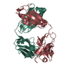
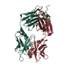
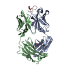
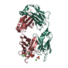
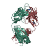
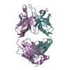
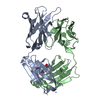
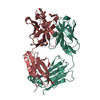
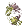
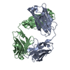
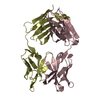
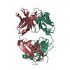
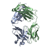
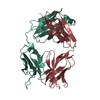
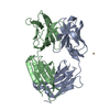
| Title | Structure of MA28-7 neutralizing antibody Fab fragment from electron cryo-microscopy of enterovirus 71 complexed with a Fab fragment | ||||||
 Components Components |
| ||||||
 Keywords Keywords | IMMUNE SYSTEM / EV71 / enterovirus / HFMD / strain-specific eptitope / VP1-145 | ||||||
| Biological species |  | ||||||
| Method | ELECTRON MICROSCOPY / single particle reconstruction / cryo EM / Resolution: 23.4 Å | ||||||
 Authors Authors | Lee, H. / Cifuente, J.O. / Ashley, R.E. / Conway, J.F. / Makhov, A.M. / Tano, Y. / Shimizu, H. / Nishimura, Y. / Hafenstein, S. | ||||||
 Citation Citation | Journal: J Virol / Year: 2013 Title: A strain-specific epitope of enterovirus 71 identified by cryo-electron microscopy of the complex with fab from neutralizing antibody. Authors: Hyunwook Lee / Javier O Cifuente / Robert E Ashley / James F Conway / Alexander M Makhov / Yoshio Tano / Hiroyuki Shimizu / Yorihiro Nishimura / Susan Hafenstein /  Abstract: Enterovirus 71 (EV71) is a picornavirus that causes outbreaks of hand, foot, and mouth disease (HFMD), primarily in the Asia-Pacific area. Unlike coxsackievirus A16, which also causes HFMD, EV71 ...Enterovirus 71 (EV71) is a picornavirus that causes outbreaks of hand, foot, and mouth disease (HFMD), primarily in the Asia-Pacific area. Unlike coxsackievirus A16, which also causes HFMD, EV71 induces severe neuropathology leading to high fatalities, especially among children under the age of 6 years. Currently, no established vaccines or treatments are available against EV71 infection. The monoclonal antibody MA28-7 neutralizes only specific strains of EV71 that have a conserved glycine at amino acid VP1-145, a surface-exposed residue that maps to the 5-fold vertex and that has been implicated in receptor binding. The cryo-electron microscopy structure of a complex between EV71 and the Fab fragment of MA28-7 shows that only one Fab fragment occupies each 5-fold vertex. A positively charged patch, which has also been implicated in receptor binding, lies within the Fab footprint. We identify the strain-specific epitope of EV71 and discuss the possible neutralization mechanisms of the antibody. | ||||||
| History |
|
- Structure visualization
| Movie |
 Movie viewer Movie viewer |
|---|---|
| Structure viewer | Molecule: MolmilJmol/JSmol |
- Downloads & links
-Download
| PDBx/mmCIF format | 3j3z.cif.gz | 92.9 KB | Display | PDBx/mmCIF format |
|---|---|---|---|---|
| PDB format | pdb3j3z.ent.gz | 69.9 KB | Display | PDB format |
| PDBx/mmJSON format | 3j3z.json.gz | Tree view | PDBx/mmJSON format | |
| Others |  Other downloads Other downloads |
-Validation report
| Arichive directory | https://data.pdbj.org/pub/pdb/validation_reports/j3/3j3zftp://data.pdbj.org/pub/pdb/validation_reports/j3/3j3z | HTTPS FTP |
|---|
-Related structure data
| Related structure data | 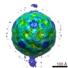 5673MC M: map data used to model this data C: citing same article ( |
|---|---|
| Similar structure data |
-Links
 PDBj
PDBj

- Assembly
| Deposited unit | 
|
|---|---|
| 1 |
|
-Components
| #1: Antibody | Mass: 23261.770 Da / Num. of mol.: 1 / Fragment: Fab / Source method: isolated from a natural source / Source: (natural) |
|---|---|
| #2: Antibody | Mass: 23349.105 Da / Num. of mol.: 1 / Fragment: Fab / Source method: isolated from a natural source / Source: (natural) |
| Has protein modification | Y |
-Experimental details
-Experiment
| Experiment | Method: ELECTRON MICROSCOPY |
|---|---|
| EM experiment | Aggregation state: PARTICLE / 3D reconstruction method: single particle reconstruction |
- Sample preparation
| Component | Name: Enterovirus 71 complexed with Fab Fragment of MA28-7 neutralizing antibody Type: VIRUS |
|---|---|
| Molecular weight | Value: 8.05 MDa / Experimental value: NO |
| Details of virus | Empty: NO / Enveloped: NO / Host category: VERTEBRATES / Isolate: STRAIN / Type: VIRION |
| Natural host | Organism: Homo sapiens |
| Buffer solution | pH: 7.5 / Details: 10 mM Tris, 200 mM NaCl, 50 mM MgCl2 |
| Specimen | Embedding applied: NO / Shadowing applied: NO / Staining applied: NO / Vitrification applied: YES |
| Specimen support | Details: glow-discharged holey carbon Quantifoil electron microscopy grids |
| Vitrification | Instrument: FEI VITROBOT MARK III / Cryogen name: OTHER / Temp: 76 K / Humidity: 95 % Details: Plunged into ethane-propane mixture (FEI VITROBOT MARK III) |
- Electron microscopy imaging
| Experimental equipment |  Model: Tecnai F20 / Image courtesy: FEI Company |
|---|---|
| Microscopy | Model: FEI TECNAI F20 / Date: Feb 2, 2011 |
| Electron gun | Electron source:  FIELD EMISSION GUN / Accelerating voltage: 200 kV / Illumination mode: SPOT SCAN FIELD EMISSION GUN / Accelerating voltage: 200 kV / Illumination mode: SPOT SCAN |
| Electron lens | Mode: BRIGHT FIELD / Nominal magnification: 50000 X / Calibrated magnification: 48425 X / Nominal defocus max: 6120 nm / Nominal defocus min: 1900 nm / Cs: 2 mm / Camera length: 0 mm |
| Specimen holder | Specimen holder model: GATAN LIQUID NITROGEN / Temperature: 95 K / Tilt angle max: 0 ° / Tilt angle min: 0 ° |
| Image recording | Electron dose: 15 e/Å2 / Film or detector model: KODAK SO-163 FILM |
| Image scans | Num. digital images: 45 |
| Radiation | Protocol: SINGLE WAVELENGTH / Monochromatic (M) / Laue (L): M / Scattering type: x-ray |
| Radiation wavelength | Relative weight: 1 |
- Processing
| EM software |
| ||||||||||||
|---|---|---|---|---|---|---|---|---|---|---|---|---|---|
| CTF correction | Details: Each | ||||||||||||
| Symmetry | Point symmetry: C1 (asymmetric) | ||||||||||||
| 3D reconstruction | Method: Cross correlation coefficient / Resolution: 23.4 Å / Resolution method: FSC 0.5 CUT-OFF / Num. of particles: 27026 / Nominal pixel size: 1.23 Å / Actual pixel size: 1.23 Å Details: The breaksym option in EMAN2 was used to search for the icosahedrally-related orientations. (Single particle details: Selected particles were low-pass filtered at 10 Angstrom. Asymmetric ...Details: The breaksym option in EMAN2 was used to search for the icosahedrally-related orientations. (Single particle details: Selected particles were low-pass filtered at 10 Angstrom. Asymmetric reconstruction was performed with the breaksym option in EMAN2.) (Single particle--Applied symmetry: C1) Num. of class averages: 2300 / Symmetry type: POINT | ||||||||||||
| Atomic model building | Protocol: RIGID BODY FIT / Space: REAL / Target criteria: correlation coefficient Details: REFINEMENT PROTOCOL--rigid body DETAILS--The Fab structure was fitted manually, then refined in Chimera. | ||||||||||||
| Atomic model building | 3D fitting-ID: 1 / Accession code: 3GK8 / Initial refinement model-ID: 1 / PDB-ID: 3GK8 / Source name: PDB / Type: experimental model
| ||||||||||||
| Refinement step | Cycle: LAST
|