 Movie
Movie Controller


+ Open data

- Basic information
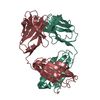
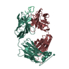
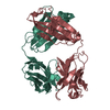
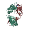
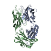
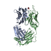
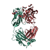
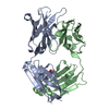
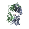
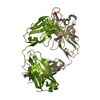
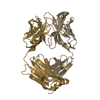
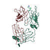
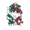
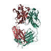
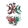
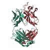
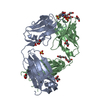
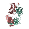
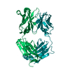
| Entry | Database: PDB / ID: 1kcs | ||||||
|---|---|---|---|---|---|---|---|
| Title | CRYSTAL STRUCTURE OF ANTIBODY PC282 IN COMPLEX WITH PS1 PEPTIDE | ||||||
 Components Components |
| ||||||
 Keywords Keywords | IMMUNE SYSTEM / ANTIBODY / PEPTIDE ANTIGEN COMPLEX (ANTIBODY-PEPTIDE) | ||||||
| Function / homology |  Function and homology information Function and homology informationcaveolin-mediated endocytosis of virus by host cell / alpha-beta T cell receptor complex / IgG immunoglobulin complex / immunoglobulin mediated immune response / immunoglobulin complex / antigen binding / B cell differentiation / adaptive immune response / fusion of virus membrane with host endosome membrane / viral envelope ...caveolin-mediated endocytosis of virus by host cell / alpha-beta T cell receptor complex / IgG immunoglobulin complex / immunoglobulin mediated immune response / immunoglobulin complex / antigen binding / B cell differentiation / adaptive immune response / fusion of virus membrane with host endosome membrane / viral envelope / virion attachment to host cell / virion membrane / extracellular region / membrane / plasma membrane Similarity search - Function | ||||||
| Biological species |  | ||||||
| Method |  X-RAY DIFFRACTION / MOLECULAR REPLACEMENT / Resolution: 2.5 Å X-RAY DIFFRACTION / MOLECULAR REPLACEMENT / Resolution: 2.5 Å | ||||||
 Authors Authors | Nair, D.T. / Singh, K. / Siddiqui, Z. / Nayak, B.P. / Rao, K.V.S. / Salunke, D.M. | ||||||
 Citation Citation | Journal: J.Immunol. / Year: 2002 Title: Epitope recognition by diverse antibodies suggests conformational convergence in an antibody response. Authors: Nair, D.T. / Singh, K. / Siddiqui, Z. / Nayak, B.P. / Rao, K.V. / Salunke, D.M. #1: Journal: J.IMMUNOL. / Year: 2000Title: Crystal Structure of an Antibody Bound to an Immunodominant Peptide Epitope: Novel Features in Peptide-antibody Recognition Authors: Nair, D.T. / Singh, K. / Sahu, N. / Rao, K.V. / Salunke, D.M. | ||||||
| History |
| ||||||
| Remark 999 | SEQUENCE An appropriate sequence database match for the Immunoglobulin was not available at the ...SEQUENCE An appropriate sequence database match for the Immunoglobulin was not available at the time of processing. |
- Structure visualization
| Structure viewer | Molecule: MolmilJmol/JSmol |
|---|
- Downloads & links
-Download
| PDBx/mmCIF format | 1kcs.cif.gz | 89.2 KB | Display | PDBx/mmCIF format |
|---|---|---|---|---|
| PDB format | pdb1kcs.ent.gz | 68.2 KB | Display | PDB format |
| PDBx/mmJSON format | 1kcs.json.gz | Tree view | PDBx/mmJSON format | |
| Others |  Other downloads Other downloads |
-Validation report
| Arichive directory | https://data.pdbj.org/pub/pdb/validation_reports/kc/1kcsftp://data.pdbj.org/pub/pdb/validation_reports/kc/1kcs | HTTPS FTP |
|---|
-Related structure data


-Links
 PDBj
PDBj


- Assembly
| Deposited unit | 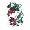
| ||||||||
|---|---|---|---|---|---|---|---|---|---|
| 1 |
| ||||||||
| Unit cell |
|
-Components
| #1: Antibody | Mass: 22983.266 Da / Num. of mol.: 1 / Fragment: light chain / Source method: isolated from a natural source / Source: (natural) |
|---|---|
| #2: Antibody | Mass: 22760.301 Da / Num. of mol.: 1 / Fragment: heavy chain / Source method: isolated from a natural source / Source: (natural) |
| #3: Protein/peptide | Mass: 1584.622 Da / Num. of mol.: 1 / Source method: obtained synthetically Details: The peptide was chemically synthesized. The sequence of the peptide is naturally found in Hepatitis B virus. References: GenBank: 15419846, UniProt: Q91C35*PLUS |
| #4: Water | ChemComp-HOH /  Mass: 18.015 Da / Num. of mol.: 85 / Source method: isolated from a natural source / Formula: H2O Mass: 18.015 Da / Num. of mol.: 85 / Source method: isolated from a natural source / Formula: H2O |
| Has protein modification | Y |
-Experimental details
-Experiment
| Experiment | Method: X-RAY DIFFRACTION / Number of used crystals: 1 |
|---|
- Sample preparation
| Crystal | Density Matthews: 2.28 Å3/Da / Density % sol: 46.08 % | ||||||||||||||||||||||||
|---|---|---|---|---|---|---|---|---|---|---|---|---|---|---|---|---|---|---|---|---|---|---|---|---|---|
| Crystal grow | Temperature: 300 K / Method: vapor diffusion, hanging drop / pH: 7.2 Details: 20% PEG 8K, pH 7.2, VAPOR DIFFUSION, HANGING DROP, temperature 300K | ||||||||||||||||||||||||
| Crystal grow | *PLUS | ||||||||||||||||||||||||
| Components of the solutions | *PLUS
|
-Data collection
| Diffraction | Mean temperature: 300 K |
|---|---|
| Diffraction source | Source: ROTATING ANODE / Type: RIGAKU / Wavelength: 1.5418 Å |
| Detector | Type: MARRESEARCH / Detector: IMAGE PLATE |
| Radiation | Monochromator: GRAPHITE / Protocol: SINGLE WAVELENGTH / Monochromatic (M) / Laue (L): M / Scattering type: x-ray |
| Radiation wavelength | Wavelength: 1.5418 Å / Relative weight: 1 |
| Reflection | Resolution: 2.5→50 Å / Num. obs: 14014 / % possible obs: 84 % / Redundancy: 4.5 % / Biso Wilson estimate: 34.9 Å2 / Rmerge(I) obs: 0.102 / Net I/σ(I): 4.9 |
| Reflection | *PLUS Highest resolution: 2.5 Å / Lowest resolution: 50 Å / Num. all: 62432 / % possible obs: 84 % |
| Reflection shell | *PLUS % possible obs: 83.2 % / Rmerge(I) obs: 0.471 / Mean I/σ(I) obs: 1.1 |
- Processing
| Software |
| |||||||||||||||||||||
|---|---|---|---|---|---|---|---|---|---|---|---|---|---|---|---|---|---|---|---|---|---|---|
| Refinement | Method to determine structure: MOLECULAR REPLACEMENT / Resolution: 2.5→50 Å / Rfactor Rfree error: 0.01 / Data cutoff high absF: 175126.26 / Data cutoff low absF: 0 / Isotropic thermal model: anisotropic / Cross valid method: THROUGHOUT / σ(F): 0 / Stereochemistry target values: ENGH & HUBER
| |||||||||||||||||||||
| Solvent computation | Solvent model: FLAT MODEL / Bsol: 46.4748 Å2 / ksol: 0.301772 e/Å3 | |||||||||||||||||||||
| Displacement parameters | Biso mean: 31.9 Å2
| |||||||||||||||||||||
| Refine analyze |
| |||||||||||||||||||||
| Refinement step | Cycle: LAST / Resolution: 2.5→50 Å
| |||||||||||||||||||||
| Refine LS restraints |
| |||||||||||||||||||||
| LS refinement shell | Resolution: 2.5→2.66 Å / Rfactor Rfree error: 0.033 / Total num. of bins used: 6
| |||||||||||||||||||||
| Xplor file |
| |||||||||||||||||||||
| Refinement | *PLUS Highest resolution: 2.5 Å / Lowest resolution: 50 Å / Rfactor obs: 0.209 | |||||||||||||||||||||
| Solvent computation | *PLUS | |||||||||||||||||||||
| Displacement parameters | *PLUS | |||||||||||||||||||||
| Refine LS restraints | *PLUS
| |||||||||||||||||||||
| LS refinement shell | *PLUS Rfactor Rfree: 0.325 / Rfactor obs: 0.309 |
