#256 - Apr 2021 SARS-CoV-2 Spike and Antibodies similarity (1)
-
Assembly
Deposited unit
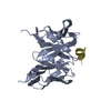
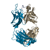
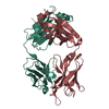
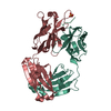
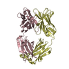
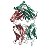
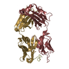
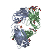
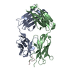
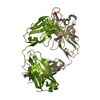
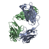
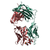
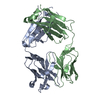
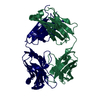
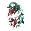
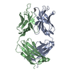
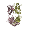
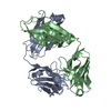
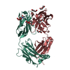
A: Genome polyprotein H: Heavy chain of Fab fragment derived from non-neutralizing antibody DAO5 L: Light chain of Fab fragment derived from non-neutralizing antibody DAO5
Protocol: SINGLE WAVELENGTH / Monochromatic (M) / Laue (L): M / Scattering type: x-ray
Radiation wavelength
Wavelength: 0.98011 Å / Relative weight: 1
Reflection
Resolution: 1.7→48.44 Å / Num. obs: 44810 / % possible obs: 97.1 % / Redundancy: 5.7 % / Biso Wilson estimate: 19.53 Å2 / Rmerge(I) obs: 0.04 / Net I/σ(I): 27.6
Reflection shell
Resolution: 1.7→1.79 Å / Redundancy: 5.7 % / Rmerge(I) obs: 0.228 / Mean I/σ(I) obs: 6.8 / Num. unique obs: 6417 / % possible all: 95.6
-
Processing
Software
Name
Version
Classification
BUSTER
2.10.2
refinement
XDS
datareduction
XSCALE
datascaling
PHASER
phasing
Refinement
Method to determine structure: MOLECULAR REPLACEMENT Starting model: Fab fragment Resolution: 1.7→34 Å / Cor.coef. Fo:Fc: 0.9355 / Cor.coef. Fo:Fc free: 0.926 / SU R Cruickshank DPI: 0.107 / Cross valid method: THROUGHOUT / σ(F): 0 / SU R Blow DPI: 0.11 / SU Rfree Blow DPI: 0.101 / SU Rfree Cruickshank DPI: 0.1
Rfactor
Num. reflection
% reflection
Selection details
Rfree
0.2025
2254
5.03 %
RANDOM
Rwork
0.1788
-
-
-
obs
0.18
44782
96.94 %
-
Displacement parameters
Biso mean: 20.06 Å2
Baniso -1
Baniso -2
Baniso -3
1-
2.993 Å2
0 Å2
1.0645 Å2
2-
-
-6.9838 Å2
0 Å2
3-
-
-
3.9909 Å2
Refine analyze
Luzzati coordinate error obs: 0.209 Å
Refinement step
Cycle: 1 / Resolution: 1.7→34 Å
Protein
Nucleic acid
Ligand
Solvent
Total
Num. atoms
3353
0
0
329
3682
Refine LS restraints
Refine-ID
Type
Dev ideal
Number
Restraint function
Weight
X-RAY DIFFRACTION
t_bond_d
0.01
3450
HARMONIC
2
X-RAY DIFFRACTION
t_angle_deg
1.1
4699
HARMONIC
2
X-RAY DIFFRACTION
t_dihedral_angle_d
1131
SINUSOIDAL
2
X-RAY DIFFRACTION
t_incorr_chiral_ct
X-RAY DIFFRACTION
t_pseud_angle
X-RAY DIFFRACTION
t_trig_c_planes
72
HARMONIC
2
X-RAY DIFFRACTION
t_gen_planes
498
HARMONIC
5
X-RAY DIFFRACTION
t_it
3450
HARMONIC
20
X-RAY DIFFRACTION
t_nbd
X-RAY DIFFRACTION
t_omega_torsion
4.36
X-RAY DIFFRACTION
t_other_torsion
15.01
X-RAY DIFFRACTION
t_improper_torsion
X-RAY DIFFRACTION
t_chiral_improper_torsion
467
SEMIHARMONIC
5
X-RAY DIFFRACTION
t_sum_occupancies
X-RAY DIFFRACTION
t_utility_distance
X-RAY DIFFRACTION
t_utility_angle
X-RAY DIFFRACTION
t_utility_torsion
X-RAY DIFFRACTION
t_ideal_dist_contact
4194
SEMIHARMONIC
4
LS refinement shell
Resolution: 1.7→1.74 Å / Total num. of bins used: 20
Rfactor
Num. reflection
% reflection
Rfree
0.2034
182
5.58 %
Rwork
0.1712
3077
-
all
0.173
3259
-
obs
-
-
95.17 %
+
About Yorodumi
-
News
-
Feb 9, 2022. New format data for meta-information of EMDB entries
New format data for meta-information of EMDB entries
Version 3 of the EMDB header file is now the official format.
The previous official version 1.9 will be removed from the archive.
In the structure databanks used in Yorodumi, some data are registered as the other names, "COVID-19 virus" and "2019-nCoV". Here are the details of the virus and the list of structure data.
Jan 31, 2019. EMDB accession codes are about to change! (news from PDBe EMDB page)
EMDB accession codes are about to change! (news from PDBe EMDB page)
The allocation of 4 digits for EMDB accession codes will soon come to an end. Whilst these codes will remain in use, new EMDB accession codes will include an additional digit and will expand incrementally as the available range of codes is exhausted. The current 4-digit format prefixed with “EMD-” (i.e. EMD-XXXX) will advance to a 5-digit format (i.e. EMD-XXXXX), and so on. It is currently estimated that the 4-digit codes will be depleted around Spring 2019, at which point the 5-digit format will come into force.
The EM Navigator/Yorodumi systems omit the EMD- prefix.
Related info.:Q: What is EMD? / ID/Accession-code notation in Yorodumi/EM Navigator
Yorodumi is a browser for structure data from EMDB, PDB, SASBDB, etc.
This page is also the successor to EM Navigator detail page, and also detail information page/front-end page for Omokage search.
The word "yorodu" (or yorozu) is an old Japanese word meaning "ten thousand". "mi" (miru) is to see.
Related info.:EMDB / PDB / SASBDB / Comparison of 3 databanks / Yorodumi Search / Aug 31, 2016. New EM Navigator & Yorodumi / Yorodumi Papers / Jmol/JSmol / Function and homology information / Changes in new EM Navigator and Yorodumi
 Movie
Movie Controller

 Yorodumi
 Open data
 Basic information
 Components
 Keywords
 Function and homology information
 Hepatitis C virus
 X-RAY DIFFRACTION /
  Authors
 Germany,
  France, 3items
  Citation
 Structure visualization
 Downloads & links
 Other downloads
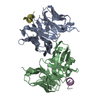


 PDBj
PDBj


 Assembly
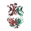

 Mass: 18.015 Da / Num. of mol.: 329 / Source method: isolated from a natural source / Formula: H2O
 Sample preparation
 Processing