 Movie
Movie Controller

[English] 日本語
 Yorodumi
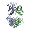
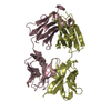
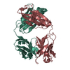
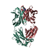
- PDB-1dqd: CRYSTAL STRUCTURE OF FAB HGR-2 F6, A COMPETITIVE ANTAGONIST OF TH... -
+ Open data

- Basic information
| Entry | Database: PDB / ID: 1dqd | ||||||
|---|---|---|---|---|---|---|---|
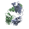
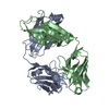
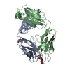
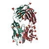
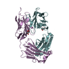
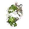
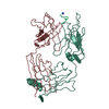
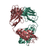
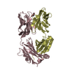
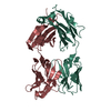
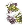
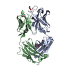
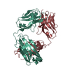
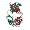
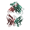
| Title | CRYSTAL STRUCTURE OF FAB HGR-2 F6, A COMPETITIVE ANTAGONIST OF THE GLUCAGON RECEPTOR | ||||||
 Components Components | (FAB HGR-2 F6) x 2 | ||||||
 Keywords Keywords | IMMUNE SYSTEM / Glucagon receptor / monoclonal antibody / Fab / receptor antagonist / typical immunoglobulin fold / light chain / heavy chain / antigen binding site / complementarity-determining regions | ||||||
| Function / homology | Immunoglobulins / Immunoglobulin-like / Sandwich / Mainly Beta Function and homology information Function and homology information | ||||||
| Biological species |  | ||||||
| Method |  X-RAY DIFFRACTION / SYNCHROTRON / Resolution: 2.1 Å X-RAY DIFFRACTION / SYNCHROTRON / Resolution: 2.1 Å | ||||||
 Authors Authors | Wright, L.M. / Brzozowski, A.M. / Hubbard, R.E. / Pike, A.C.W. / Roberts, S.M. / Skovgaard, R.N. / Svendsen, I. / Vissing, H. / Bywater, R.P. | ||||||
 Citation Citation | Journal: Acta Crystallogr.,Sect.D / Year: 2000 Title: Structure of Fab hGR-2 F6, a competitive antagonist of the glucagon receptor. Authors: Wright, L.M. / Brzozowski, A.M. / Hubbard, R.E. / Pike, A.C. / Roberts, S.M. / Skovgaard, R.N. / Svendsen, I. / Vissing, H. / Bywater, R.P. | ||||||
| History |
|
- Structure visualization
| Structure viewer | Molecule: MolmilJmol/JSmol |
|---|
- Downloads & links
-Download
| PDBx/mmCIF format | 1dqd.cif.gz | 105.6 KB | Display | PDBx/mmCIF format |
|---|---|---|---|---|
| PDB format | pdb1dqd.ent.gz | 80.6 KB | Display | PDB format |
| PDBx/mmJSON format | 1dqd.json.gz | Tree view | PDBx/mmJSON format | |
| Others |  Other downloads Other downloads |
-Validation report
| Arichive directory | https://data.pdbj.org/pub/pdb/validation_reports/dq/1dqdftp://data.pdbj.org/pub/pdb/validation_reports/dq/1dqd | HTTPS FTP |
|---|
-Related structure data
| Similar structure data |
|---|

-Links
 PDBj
PDBj

- Assembly
| Deposited unit | 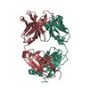
| ||||||||
|---|---|---|---|---|---|---|---|---|---|
| 1 |
| ||||||||
| Unit cell |
| ||||||||
| Details | The light chain and the heavy chain hydrogen bond together to form a single Fab monomer within the asymmetric unit. |
-Components
| #1: Antibody | Mass: 23953.500 Da / Num. of mol.: 1 / Fragment: LIGHT CHAIN / Source method: isolated from a natural source Details: Using RBF mice, monoclonal antibodies were generated against the intact Human membrane-bound glucagon receptor. Fab fragments were produced from the intact IgG monoclonal antibody by papain ...Details: Using RBF mice, monoclonal antibodies were generated against the intact Human membrane-bound glucagon receptor. Fab fragments were produced from the intact IgG monoclonal antibody by papain digestion. Final purification of the Fab fragments was performed using both a MonoQ ion exchange column and subsequently a MonoS column. Source: (natural) |
|---|---|
| #2: Antibody | Mass: 23892.545 Da / Num. of mol.: 1 / Fragment: HEAVY CHAIN / Source method: isolated from a natural source Details: Using RBF mice, monoclonal antibodies were generated against the intact Human membrane-bound glucagon receptor. Fab fragments were produced from the intact IgG monoclonal antibody by papain ...Details: Using RBF mice, monoclonal antibodies were generated against the intact Human membrane-bound glucagon receptor. Fab fragments were produced from the intact IgG monoclonal antibody by papain digestion. Final purification of the Fab fragments was performed using both a MonoQ ion exchange column and subsequently a MonoS column. Source: (natural) |
| #3: Water | ChemComp-HOH /  Mass: 18.015 Da / Num. of mol.: 380 / Fragment: WATER / Source method: isolated from a natural source / Formula: H2O Mass: 18.015 Da / Num. of mol.: 380 / Fragment: WATER / Source method: isolated from a natural source / Formula: H2O |
| Has protein modification | Y |
-Experimental details
-Experiment
| Experiment | Method: X-RAY DIFFRACTION / Number of used crystals: 1 |
|---|
- Sample preparation
| Crystal | Density Matthews: 1.99 Å3/Da / Density % sol: 38.25 % | |||||||||||||||||||||||||
|---|---|---|---|---|---|---|---|---|---|---|---|---|---|---|---|---|---|---|---|---|---|---|---|---|---|---|
| Crystal grow | Temperature: 291 K / Method: vapor diffusion, sitting drop / pH: 8.5 Details: 24% (w/w) PEG 2KME, 100mM Tris HCl , pH 8.5, VAPOR DIFFUSION, SITTING DROP, temperature 291.0K | |||||||||||||||||||||||||
| Crystal grow | *PLUS pH: 8 | |||||||||||||||||||||||||
| Components of the solutions | *PLUS
|
-Data collection
| Diffraction | Mean temperature: 100 K |
|---|---|
| Diffraction source | Source: SYNCHROTRON / Site: SRS  / Beamline: PX9.5 / Wavelength: 0.92 / Beamline: PX9.5 / Wavelength: 0.92 |
| Detector | Type: MARRESEARCH / Detector: IMAGE PLATE / Date: Jul 1, 1998 |
| Radiation | Protocol: SINGLE WAVELENGTH / Monochromatic (M) / Laue (L): M / Scattering type: x-ray |
| Radiation wavelength | Wavelength: 0.92 Å / Relative weight: 1 |
| Reflection | Resolution: 2.1→20 Å / Num. all: 173940 / Num. obs: 22946 / % possible obs: 98.1 % / Redundancy: 4 % / Biso Wilson estimate: 33.1 Å2 / Rmerge(I) obs: 0.053 / Net I/σ(I): 10.4 |
| Reflection shell | Resolution: 2.1→2.14 Å / Rmerge(I) obs: 0.418 / Num. unique all: 1061 / % possible all: 95.4 |
| Reflection | *PLUS Num. measured all: 173940 |
| Reflection shell | *PLUS % possible obs: 95.4 % / Rmerge(I) obs: 0.319 / Mean I/σ(I) obs: 2.6 |
- Processing
| Software |
| |||||||||||||||||||||||||
|---|---|---|---|---|---|---|---|---|---|---|---|---|---|---|---|---|---|---|---|---|---|---|---|---|---|---|
| Refinement | Resolution: 2.1→20 Å / σ(F): 0 / σ(I): 0 / Stereochemistry target values: Engh & Huber / Details: Used maximum likelihood method.
| |||||||||||||||||||||||||
| Refinement step | Cycle: LAST / Resolution: 2.1→20 Å
| |||||||||||||||||||||||||
| Software | *PLUS Name: REFMAC / Classification: refinement | |||||||||||||||||||||||||
| Refinement | *PLUS Highest resolution: 2.1 Å / σ(F): 0 / % reflection Rfree: 5 % / Rfactor obs: 0.217 | |||||||||||||||||||||||||
| Solvent computation | *PLUS | |||||||||||||||||||||||||
| Displacement parameters | *PLUS | |||||||||||||||||||||||||
| Refine LS restraints | *PLUS
|
