 Movie
Movie Controller

[English] 日本語
 Yorodumi
- PDB-1y18: Fab fragment of catalytic elimination antibody 34E4 E(H50)D mutan... -
+ Open data

- Basic information
| Entry | Database: PDB / ID: 1y18 | ||||||
|---|---|---|---|---|---|---|---|
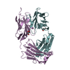
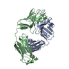
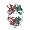
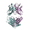
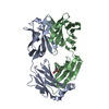
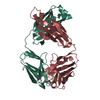
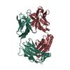
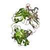
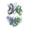
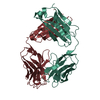
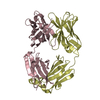
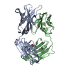
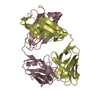
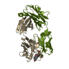
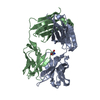
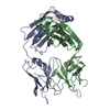
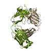
| Title | Fab fragment of catalytic elimination antibody 34E4 E(H50)D mutant in complex with hapten | ||||||
 Components Components |
| ||||||
 Keywords Keywords | IMMUNE SYSTEM / Immunoglobulin / Catalytic antibody / chimeric Fab / Hapten complex | ||||||
| Function / homology | Immunoglobulins / Immunoglobulin-like / Sandwich / Mainly Beta / Chem-HAN Function and homology information Function and homology information | ||||||
| Biological species |   Homo sapiens (human) Homo sapiens (human) | ||||||
| Method |  X-RAY DIFFRACTION / SYNCHROTRON / MOLECULAR REPLACEMENT / Resolution: 2.8 Å X-RAY DIFFRACTION / SYNCHROTRON / MOLECULAR REPLACEMENT / Resolution: 2.8 Å | ||||||
 Authors Authors | Debler, E.W. / Ito, S. / Heine, A. / Wilson, I.A. | ||||||
 Citation Citation | Journal: Proc.Natl.Acad.Sci.USA / Year: 2005 Title: Structural origins of efficient proton abstraction from carbon by a catalytic antibody Authors: Debler, E.W. / Ito, S. / Seebeck, F.P. / Heine, A. / Hilvert, D. / Wilson, I.A. | ||||||
| History |
| ||||||
| Remark 999 | SEQUENCE THE SEQUENCES OF THE FAB COMPLEXES ARE NOT YET AVAILABLE IN ANY REFERENCE SEQUENCE ...SEQUENCE THE SEQUENCES OF THE FAB COMPLEXES ARE NOT YET AVAILABLE IN ANY REFERENCE SEQUENCE DATABASES. RESIDUE 50 IN CHAINS H,B,D and F WAS MUTATED FROM GLU TO ASP. |
- Structure visualization
| Structure viewer | Molecule: MolmilJmol/JSmol |
|---|
- Downloads & links
-Download
| PDBx/mmCIF format | 1y18.cif.gz | 342.5 KB | Display | PDBx/mmCIF format |
|---|---|---|---|---|
| PDB format | pdb1y18.ent.gz | 278.9 KB | Display | PDB format |
| PDBx/mmJSON format | 1y18.json.gz | Tree view | PDBx/mmJSON format | |
| Others |  Other downloads Other downloads |
-Validation report
| Arichive directory | https://data.pdbj.org/pub/pdb/validation_reports/y1/1y18ftp://data.pdbj.org/pub/pdb/validation_reports/y1/1y18 | HTTPS FTP |
|---|
-Related structure data
| Related structure data |  1y0lSC S: Starting model for refinement C: citing same article ( |
|---|---|
| Similar structure data |


-Links
 PDBj
PDBj

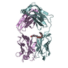
- Assembly
| Deposited unit | 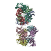
| ||||||||
|---|---|---|---|---|---|---|---|---|---|
| 1 | 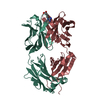
| ||||||||
| 2 | 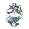
| ||||||||
| 3 | 
| ||||||||
| 4 | 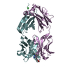
| ||||||||
| Unit cell |
|
-Components
| #1: Antibody | Mass: 23258.826 Da / Num. of mol.: 4 / Mutation: E(H50)D Source method: isolated from a genetically manipulated source Details: The variable domain (residues 1-107) is from a murine source and the constant domain (108-214) is from a human source Source: (gene. exp.) Mus musculus, Homo sapiens / Genus: Mus, Homo / Species: , / Strain: , / Plasmid: p4xH-M13 / Production host:  #2: Antibody | Mass: 24572.699 Da / Num. of mol.: 4 / Mutation: E(H50)D Source method: isolated from a genetically manipulated source Details: The variable domain (residues 1-113) is from a murine source and the constant domain (108-233) is from a human source Source: (gene. exp.) Mus musculus, Homo sapiens / Genus: Mus, Homo / Species: , / Strain: , / Plasmid: p4xH-M13 / Production host: #3: Chemical | ChemComp-HAN /   Mass: 261.320 Da / Num. of mol.: 4 / Source method: obtained synthetically / Formula: C14H19N3O2 Mass: 261.320 Da / Num. of mol.: 4 / Source method: obtained synthetically / Formula: C14H19N3O2#4: Chemical | ChemComp-CL / |   Mass: 35.453 Da / Num. of mol.: 1 / Source method: obtained synthetically / Formula: Cl Mass: 35.453 Da / Num. of mol.: 1 / Source method: obtained synthetically / Formula: Cl#5: Water | ChemComp-HOH / |  Mass: 18.015 Da / Num. of mol.: 287 / Source method: isolated from a natural source / Formula: H2O Mass: 18.015 Da / Num. of mol.: 287 / Source method: isolated from a natural source / Formula: H2OHas protein modification | Y | |
|---|
-Experimental details
-Experiment
| Experiment | Method: X-RAY DIFFRACTION / Number of used crystals: 1 |
|---|
- Sample preparation
| Crystal | Density Matthews: 3 Å3/Da / Density % sol: 57 % |
|---|---|
| Crystal grow | Method: vapor diffusion, sitting drop / pH: 8 Details: Diammonium hydrogen phosphate, NaCl, imidazole HCl, pH 8.0, VAPOR DIFFUSION, SITTING DROP, temperature 110K |
-Data collection
| Diffraction | Mean temperature: 100 K |
|---|---|
| Diffraction source | Source: SYNCHROTRON / Site: ALS  / Beamline: 8.3.1 / Wavelength: 1.11587 Å / Beamline: 8.3.1 / Wavelength: 1.11587 Å |
| Detector | Type: ADSC QUANTUM 210 / Detector: CCD / Date: Oct 10, 2003 |
| Radiation | Monochromator: Double Crystal / Protocol: SINGLE WAVELENGTH / Monochromatic (M) / Laue (L): M / Scattering type: x-ray |
| Radiation wavelength | Wavelength: 1.11587 Å / Relative weight: 1 |
| Reflection | Resolution: 2.8→50 Å / Num. all: 57674 / Num. obs: 56867 / % possible obs: 98.6 % / Observed criterion σ(F): 0 / Observed criterion σ(I): 0 / Redundancy: 2.4 % / Biso Wilson estimate: 33.1 Å2 / Rsym value: 0.089 / Net I/σ(I): 8.2 |
| Reflection shell | Resolution: 2.8→2.85 Å / Redundancy: 2.4 % / Mean I/σ(I) obs: 1.5 / Num. unique all: 2795 / Rsym value: 0.359 / % possible all: 98.5 |
- Processing
| Software |
| |||||||||||||||||||||||||
|---|---|---|---|---|---|---|---|---|---|---|---|---|---|---|---|---|---|---|---|---|---|---|---|---|---|---|
| Refinement | Method to determine structure: MOLECULAR REPLACEMENT Starting model: pdb entry 1Y0L Resolution: 2.8→50 Å / Isotropic thermal model: ANISOTROPIC / Cross valid method: THROUGHOUT / σ(F): 1 / Stereochemistry target values: Engh & Huber
| |||||||||||||||||||||||||
| Displacement parameters | Biso mean: 32 Å2
| |||||||||||||||||||||||||
| Refine analyze |
| |||||||||||||||||||||||||
| Refinement step | Cycle: LAST / Resolution: 2.8→50 Å
| |||||||||||||||||||||||||
| Refine LS restraints |
| |||||||||||||||||||||||||
| LS refinement shell | Resolution: 2.8→2.98 Å / Rfactor Rfree error: 0.017
|
