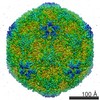
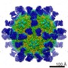
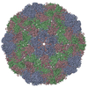
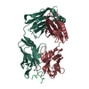
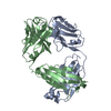
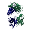
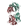
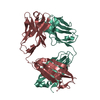
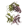
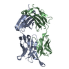
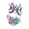
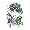
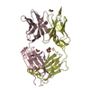
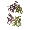
Journal: Proc Natl Acad Sci U S A / Year: 2017 Title: Potent neutralization of hepatitis A virus reveals a receptor mimic mechanism and the receptor recognition site. Authors: Xiangxi Wang / Ling Zhu / Minghao Dang / Zhongyu Hu / Qiang Gao / Shuai Yuan / Yao Sun / Bo Zhang / Jingshan Ren / Abhay Kotecha / Thomas S Walter / Junzhi Wang / Elizabeth E Fry / David I Stuart / Zihe Rao / Abstract: Hepatitis A virus (HAV) infects ∼1.4 million people annually and, although there is a vaccine, there are no licensed therapeutic drugs. HAV is unusually stable (making disinfection problematic) and ...Hepatitis A virus (HAV) infects ∼1.4 million people annually and, although there is a vaccine, there are no licensed therapeutic drugs. HAV is unusually stable (making disinfection problematic) and little is known of how it enters cells and releases its RNA. Here we report a potent HAV-specific monoclonal antibody, R10, which neutralizes HAV infection by blocking attachment to the host cell. High-resolution cryo-EM structures of HAV full and empty particles and of the complex of HAV with R10 Fab reveal the atomic details of antibody binding and point to a receptor recognition site at the pentamer interface. These results, together with our observation that the R10 Fab destabilizes the capsid, suggest the use of a receptor mimic mechanism to neutralize virus infection, providing new opportunities for therapeutic intervention.
In the structure databanks used in Yorodumi, some data are registered as the other names, "COVID-19 virus" and "2019-nCoV". Here are the details of the virus and the list of structure data.
Jan 31, 2019. EMDB accession codes are about to change! (news from PDBe EMDB page)
EMDB accession codes are about to change! (news from PDBe EMDB page)
The allocation of 4 digits for EMDB accession codes will soon come to an end. Whilst these codes will remain in use, new EMDB accession codes will include an additional digit and will expand incrementally as the available range of codes is exhausted. The current 4-digit format prefixed with “EMD-” (i.e. EMD-XXXX) will advance to a 5-digit format (i.e. EMD-XXXXX), and so on. It is currently estimated that the 4-digit codes will be depleted around Spring 2019, at which point the 5-digit format will come into force.
The EM Navigator/Yorodumi systems omit the EMD- prefix.
Related info.:Q: What is EMD? / ID/Accession-code notation in Yorodumi/EM Navigator
Yorodumi is a browser for structure data from EMDB, PDB, SASBDB, etc.
This page is also the successor to EM Navigator detail page, and also detail information page/front-end page for Omokage search.
The word "yorodu" (or yorozu) is an old Japanese word meaning "ten thousand". "mi" (miru) is to see.
Related info.:EMDB / PDB / SASBDB / Comparison of 3 databanks / Yorodumi Search / Aug 31, 2016. New EM Navigator & Yorodumi / Yorodumi Papers / Jmol/JSmol / Function and homology information / Changes in new EM Navigator and Yorodumi
 Movie
Movie Controller


 Open data
 Basic information
 Components
 Keywords
 Function and homology information
 X-RAY DIFFRACTION /
  Authors
 China, 1items
  Citation
 Structure visualization
 Downloads & links
 Other downloads
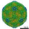


 PDBj
PDBj

 Assembly

 Sample preparation
 Processing