 Movie
Movie Controller


+ Open data

- Basic information
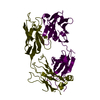
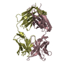
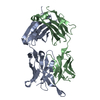
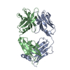
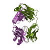
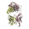
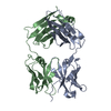
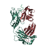
| Entry | Database: PDB / ID: 2adg | ||||||
|---|---|---|---|---|---|---|---|
| Title | Crystal structure of monoclonal anti-CD4 antibody Q425 | ||||||
 Components Components |
| ||||||
 Keywords Keywords | IMMUNE SYSTEM / anti-CD4 / interfacial metal / antibody recognition | ||||||
| Function / homology | Immunoglobulins / Immunoglobulin-like / Sandwich / Mainly Beta Function and homology information Function and homology information | ||||||
| Biological species |  | ||||||
| Method |  X-RAY DIFFRACTION / MOLECULAR REPLACEMENT / Resolution: 2.5 Å X-RAY DIFFRACTION / MOLECULAR REPLACEMENT / Resolution: 2.5 Å | ||||||
 Authors Authors | Zhou, T. / Hamer, D.H. / Hendrickson, W.A. / Sattentau, Q.J. / Kwong, P.D. | ||||||
 Citation Citation | Journal: Proc.Natl.Acad.Sci.Usa / Year: 2005 Title: Interfacial metal and antibody recognition. Authors: Zhou, T. / Hamer, D.H. / Hendrickson, W.A. / Sattentau, Q.J. / Kwong, P.D. | ||||||
| History |
| ||||||
| Remark 999 | SEQUENCE The sequence of FAB protein is not available at GB or SWS sequence database at the time of ...SEQUENCE The sequence of FAB protein is not available at GB or SWS sequence database at the time of processing. |
- Structure visualization
| Structure viewer | Molecule: MolmilJmol/JSmol |
|---|
- Downloads & links
-Download
| PDBx/mmCIF format | 2adg.cif.gz | 94.1 KB | Display | PDBx/mmCIF format |
|---|---|---|---|---|
| PDB format | pdb2adg.ent.gz | 71.3 KB | Display | PDB format |
| PDBx/mmJSON format | 2adg.json.gz | Tree view | PDBx/mmJSON format | |
| Others |  Other downloads Other downloads |
-Validation report
| Arichive directory | https://data.pdbj.org/pub/pdb/validation_reports/ad/2adgftp://data.pdbj.org/pub/pdb/validation_reports/ad/2adg | HTTPS FTP |
|---|
-Related structure data
| Related structure data |  2adiC 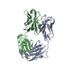 2adjC 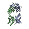 1fvdS C: citing same article ( S: Starting model for refinement |
|---|---|
| Similar structure data |


-Links
 PDBj
PDBj

- Assembly
| Deposited unit | 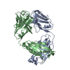
| ||||||||
|---|---|---|---|---|---|---|---|---|---|
| 1 |
| ||||||||
| Unit cell |
| ||||||||
| Details | Heterodimer of heavy and light chain |
-Components
| #1: Antibody | Mass: 23721.117 Da / Num. of mol.: 1 / Source method: isolated from a natural source / Details: Hybridoma / Source: (natural) |
|---|---|
| #2: Antibody | Mass: 23929.635 Da / Num. of mol.: 1 / Source method: isolated from a natural source / Details: Hybridoma / Source: (natural) |
| #3: Water | ChemComp-HOH /  Mass: 18.015 Da / Num. of mol.: 67 / Source method: isolated from a natural source / Formula: H2O Mass: 18.015 Da / Num. of mol.: 67 / Source method: isolated from a natural source / Formula: H2O |
| Has protein modification | Y |
-Experimental details
-Experiment
| Experiment | Method: X-RAY DIFFRACTION / Number of used crystals: 1 |
|---|
- Sample preparation
| Crystal | Density Matthews: 2.737 Å3/Da / Density % sol: 55.1 % |
|---|---|
| Crystal grow | Temperature: 298 K / Method: vapor diffusion, hanging drop / pH: 5.5 Details: PEG4000, Sodium Acetate, EDTA, pH 5.5, VAPOR DIFFUSION, HANGING DROP, temperature 298.0K |
-Data collection
| Diffraction | Mean temperature: 298 K |
|---|---|
| Diffraction source | Source: ROTATING ANODE / Type: RIGAKU / Wavelength: 1.5418 Å |
| Detector | Type: XUONG-HAMLIN MULTIWIRE / Detector: AREA DETECTOR / Date: Jan 20, 1994 |
| Radiation | Protocol: SINGLE WAVELENGTH / Monochromatic (M) / Laue (L): M / Scattering type: x-ray |
| Radiation wavelength | Wavelength: 1.5418 Å / Relative weight: 1 |
| Reflection | Resolution: 2.5→10 Å / Num. all: 18319 / Num. obs: 18319 / % possible obs: 94.4 % / Observed criterion σ(F): 2 / Observed criterion σ(I): 2 / Redundancy: 3.8 % / Biso Wilson estimate: 15.1 Å2 / Rsym value: 0.098 / Net I/σ(I): 7.3 |
| Reflection shell | Resolution: 2.5→2.61 Å / % possible all: 78.8 |
- Processing
| Software |
| ||||||||||||||||||||||||||||||||||||
|---|---|---|---|---|---|---|---|---|---|---|---|---|---|---|---|---|---|---|---|---|---|---|---|---|---|---|---|---|---|---|---|---|---|---|---|---|---|
| Refinement | Method to determine structure: MOLECULAR REPLACEMENT Starting model: pdb entry 1FVD Resolution: 2.5→10 Å / Rfactor Rfree error: 0.008 / Data cutoff high absF: 62726.54 / Data cutoff low absF: 0 / Isotropic thermal model: RESTRAINED / Cross valid method: THROUGHOUT / σ(F): 0 / Stereochemistry target values: Engh & Huber
| ||||||||||||||||||||||||||||||||||||
| Solvent computation | Solvent model: FLAT MODEL / Bsol: 30.361 Å2 / ksol: 0.316614 e/Å3 | ||||||||||||||||||||||||||||||||||||
| Displacement parameters | Biso mean: 31 Å2
| ||||||||||||||||||||||||||||||||||||
| Refine analyze |
| ||||||||||||||||||||||||||||||||||||
| Refinement step | Cycle: LAST / Resolution: 2.5→10 Å
| ||||||||||||||||||||||||||||||||||||
| Refine LS restraints |
| ||||||||||||||||||||||||||||||||||||
| LS refinement shell | Resolution: 2.5→2.66 Å / Rfactor Rfree error: 0.03 / Total num. of bins used: 6
| ||||||||||||||||||||||||||||||||||||
| Xplor file |
|
