 Movie
Movie Controller

[English] 日本語
 Yorodumi
- PDB-6y54: Crystal structure of a Neisseria meningitidis serogroup A capsula... -
+ Open data

- Basic information
| Entry | Database: PDB / ID: 6y54 | ||||||
|---|---|---|---|---|---|---|---|
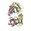
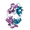
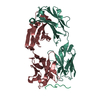
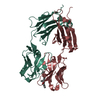
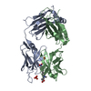
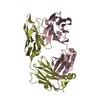
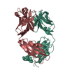
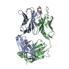
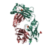
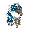
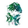
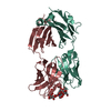
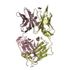
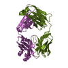
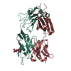
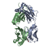
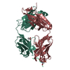
| Title | Crystal structure of a Neisseria meningitidis serogroup A capsular oligosaccharide bound to a functional Fab | ||||||
 Components Components |
| ||||||
 Keywords Keywords | IMMUNE SYSTEM / antigen-antibody complex / epitope mapping / carbohydrate / glycan | ||||||
| Function / homology | Immunoglobulins / Immunoglobulin-like / Sandwich / Mainly Beta / IODIDE ION / Chem-OA8 / Chem-OAB / Chem-OOW Function and homology information Function and homology information | ||||||
| Biological species |  | ||||||
| Method |  X-RAY DIFFRACTION / SYNCHROTRON / MOLECULAR REPLACEMENT / Resolution: 2.67 Å X-RAY DIFFRACTION / SYNCHROTRON / MOLECULAR REPLACEMENT / Resolution: 2.67 Å | ||||||
 Authors Authors | Dello Iacono, L. / Henriques, P. / Adamo, R. | ||||||
| Funding support |  Italy, 1items Italy, 1items
| ||||||
 Citation Citation | Journal: Proc.Natl.Acad.Sci.USA / Year: 2020 Title: Structure of a protective epitope reveals the importance of acetylation of Neisseria meningitidis serogroup A capsular polysaccharide. Authors: Henriques, P. / Dello Iacono, L. / Gimeno, A. / Biolchi, A. / Romano, M.R. / Arda, A. / Bernardes, G.J.L. / Jimenez-Barbero, J. / Berti, F. / Rappuoli, R. / Adamo, R. | ||||||
| History |
|
- Structure visualization
| Structure viewer | Molecule: MolmilJmol/JSmol |
|---|
- Downloads & links
-Download
| PDBx/mmCIF format | 6y54.cif.gz | 230.6 KB | Display | PDBx/mmCIF format |
|---|---|---|---|---|
| PDB format | pdb6y54.ent.gz | 180 KB | Display | PDB format |
| PDBx/mmJSON format | 6y54.json.gz | Tree view | PDBx/mmJSON format | |
| Others |  Other downloads Other downloads |
-Validation report
| Arichive directory | https://data.pdbj.org/pub/pdb/validation_reports/y5/6y54ftp://data.pdbj.org/pub/pdb/validation_reports/y5/6y54 | HTTPS FTP |
|---|
-Related structure data


-Links
 PDBj
PDBj

- Assembly
| Deposited unit | 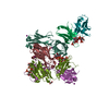
| ||||||||||||
|---|---|---|---|---|---|---|---|---|---|---|---|---|---|
| 1 | 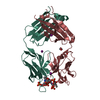
| ||||||||||||
| 2 | 
| ||||||||||||
| 3 | 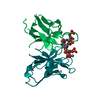
| ||||||||||||
| Unit cell |
|
-Components
-Antibody , 2 types, 6 molecules HMOLNP
| #1: Antibody | Mass: 24520.445 Da / Num. of mol.: 3 Source method: isolated from a genetically manipulated source Source: (gene. exp.)  Homo sapiens (human) Homo sapiens (human)#2: Antibody | Mass: 24225.871 Da / Num. of mol.: 3 Source method: isolated from a genetically manipulated source Source: (gene. exp.) Homo sapiens (human) |
|---|
-Non-polymers , 6 types, 47 molecules 


| #3: Chemical | ChemComp-IOD /  Mass: 126.904 Da / Num. of mol.: 9 / Source method: obtained synthetically / Formula: I Mass: 126.904 Da / Num. of mol.: 9 / Source method: obtained synthetically / Formula: I#4: Chemical |  Mass: 62.068 Da / Num. of mol.: 3 / Source method: obtained synthetically / Formula: C2H6O2 Mass: 62.068 Da / Num. of mol.: 3 / Source method: obtained synthetically / Formula: C2H6O2#5: Chemical |  Mass: 423.204 Da / Num. of mol.: 3 / Source method: obtained synthetically / Formula: C10H19NO13P2 Mass: 423.204 Da / Num. of mol.: 3 / Source method: obtained synthetically / Formula: C10H19NO13P2#6: Chemical |  Mass: 343.224 Da / Num. of mol.: 3 / Source method: obtained synthetically / Formula: C10H18NO10P Mass: 343.224 Da / Num. of mol.: 3 / Source method: obtained synthetically / Formula: C10H18NO10P#7: Chemical |  Mass: 343.224 Da / Num. of mol.: 3 / Source method: obtained synthetically / Formula: C10H18NO10P Mass: 343.224 Da / Num. of mol.: 3 / Source method: obtained synthetically / Formula: C10H18NO10P#8: Water | ChemComp-HOH / | Mass: 18.015 Da / Num. of mol.: 26 / Source method: isolated from a natural source / Formula: H2O |
|---|
-Details
| Has ligand of interest | Y |
|---|---|
| Has protein modification | Y |
-Experimental details
-Experiment
| Experiment | Method: X-RAY DIFFRACTION / Number of used crystals: 1 |
|---|
- Sample preparation
| Crystal | Density Matthews: 2.78 Å3/Da / Density % sol: 55.81 % |
|---|---|
| Crystal grow | Temperature: 293 K / Method: vapor diffusion, sitting drop / Details: 100 mM MES pH 5.4, 255 mM KI 25% PEG 4000 |
-Data collection
| Diffraction | Mean temperature: 100 K / Serial crystal experiment: N |
|---|---|
| Diffraction source | Source: SYNCHROTRON / Site: ESRF  / Beamline: ID30B / Wavelength: 0.9763 Å / Beamline: ID30B / Wavelength: 0.9763 Å |
| Detector | Type: DECTRIS PILATUS 6M / Detector: PIXEL / Date: Nov 19, 2018 |
| Radiation | Protocol: SINGLE WAVELENGTH / Monochromatic (M) / Laue (L): M / Scattering type: x-ray |
| Radiation wavelength | Wavelength: 0.9763 Å / Relative weight: 1 |
| Reflection | Resolution: 2.67→49.74 Å / Num. obs: 44254 / % possible obs: 97.6 % / Redundancy: 3.1 % / Biso Wilson estimate: 44.08 Å2 / CC1/2: 0.979 / Net I/σ(I): 6.4 |
| Reflection shell | Resolution: 2.67→2.76 Å / Num. unique obs: 4231 / CC1/2: 0.598 |
- Processing
| Software |
| |||||||||||||||||||||||||||||||||||||||||||||||||||||||||||||||||||||||||||||||||||||||||||||||||||||||||||||||||||||||
|---|---|---|---|---|---|---|---|---|---|---|---|---|---|---|---|---|---|---|---|---|---|---|---|---|---|---|---|---|---|---|---|---|---|---|---|---|---|---|---|---|---|---|---|---|---|---|---|---|---|---|---|---|---|---|---|---|---|---|---|---|---|---|---|---|---|---|---|---|---|---|---|---|---|---|---|---|---|---|---|---|---|---|---|---|---|---|---|---|---|---|---|---|---|---|---|---|---|---|---|---|---|---|---|---|---|---|---|---|---|---|---|---|---|---|---|---|---|---|---|---|
| Refinement | Method to determine structure: MOLECULAR REPLACEMENT Starting model: 5KVF, 3FO9 Resolution: 2.67→49.74 Å / SU ML: 0.4614 / Cross valid method: FREE R-VALUE / σ(F): 1.34 / Phase error: 30.4224
| |||||||||||||||||||||||||||||||||||||||||||||||||||||||||||||||||||||||||||||||||||||||||||||||||||||||||||||||||||||||
| Solvent computation | Shrinkage radii: 0.9 Å / VDW probe radii: 1.11 Å | |||||||||||||||||||||||||||||||||||||||||||||||||||||||||||||||||||||||||||||||||||||||||||||||||||||||||||||||||||||||
| Displacement parameters | Biso mean: 48.93 Å2 | |||||||||||||||||||||||||||||||||||||||||||||||||||||||||||||||||||||||||||||||||||||||||||||||||||||||||||||||||||||||
| Refinement step | Cycle: LAST / Resolution: 2.67→49.74 Å
| |||||||||||||||||||||||||||||||||||||||||||||||||||||||||||||||||||||||||||||||||||||||||||||||||||||||||||||||||||||||
| Refine LS restraints |
| |||||||||||||||||||||||||||||||||||||||||||||||||||||||||||||||||||||||||||||||||||||||||||||||||||||||||||||||||||||||
| LS refinement shell |
|
