 Movie
Movie Controller

[English] 日本語
 Yorodumi
- PDB-1nak: IGG1 FAB FRAGMENT (83.1) COMPLEX WITH 16-RESIDUE PEPTIDE (RESIDUE... -
+ Open data

- Basic information
| Entry | Database: PDB / ID: 1nak | ||||||
|---|---|---|---|---|---|---|---|
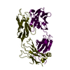
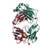
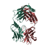
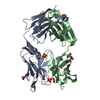
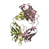
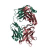
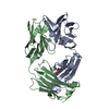
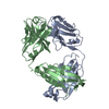
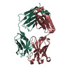
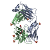
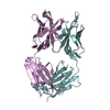
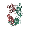
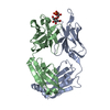
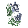
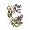
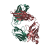
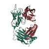
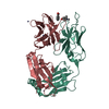
| Title | IGG1 FAB FRAGMENT (83.1) COMPLEX WITH 16-RESIDUE PEPTIDE (RESIDUES 304-321 OF HIV-1 GP120 (MN ISOLATE)) | ||||||
 Components Components |
| ||||||
 Keywords Keywords | IMMUNE SYSTEM / immunoglobulin fold | ||||||
| Function / homology |  Function and homology information Function and homology informationDectin-2 family / symbiont-mediated perturbation of host defense response / positive regulation of plasma membrane raft polarization / positive regulation of receptor clustering / host cell endosome membrane / clathrin-dependent endocytosis of virus by host cell / viral protein processing / fusion of virus membrane with host plasma membrane / fusion of virus membrane with host endosome membrane / viral envelope ...Dectin-2 family / symbiont-mediated perturbation of host defense response / positive regulation of plasma membrane raft polarization / positive regulation of receptor clustering / host cell endosome membrane / clathrin-dependent endocytosis of virus by host cell / viral protein processing / fusion of virus membrane with host plasma membrane / fusion of virus membrane with host endosome membrane / viral envelope / virion attachment to host cell / host cell plasma membrane / virion membrane / structural molecule activity / membrane / identical protein binding Similarity search - Function | ||||||
| Biological species |  | ||||||
| Method |  X-RAY DIFFRACTION / SYNCHROTRON / MOLECULAR REPLACEMENT / Resolution: 2.57 Å X-RAY DIFFRACTION / SYNCHROTRON / MOLECULAR REPLACEMENT / Resolution: 2.57 Å | ||||||
 Authors Authors | Stanfield, R.L. / Ghiara, J.B. / Saphire, E.O. / Profy, A.T. / Wilson, I.A. | ||||||
 Citation Citation | Journal: Virology / Year: 2003 Title: Recurring conformation of the human immunodeficiency virus type 1 gp120 V3 loop. Authors: Stanfield, R.L. / Ghiara, J.B. / Ollmann Saphire, E. / Profy, A.T. / Wilson, I.A. | ||||||
| History |
| ||||||
| Remark 999 | SEQUENCE The sequence of this protein was not deposited into any sequence database |
- Structure visualization
| Structure viewer | Molecule: MolmilJmol/JSmol |
|---|
- Downloads & links
-Download
| PDBx/mmCIF format | 1nak.cif.gz | 178.6 KB | Display | PDBx/mmCIF format |
|---|---|---|---|---|
| PDB format | pdb1nak.ent.gz | 142.3 KB | Display | PDB format |
| PDBx/mmJSON format | 1nak.json.gz | Tree view | PDBx/mmJSON format | |
| Others |  Other downloads Other downloads |
-Validation report
| Arichive directory | https://data.pdbj.org/pub/pdb/validation_reports/na/1nakftp://data.pdbj.org/pub/pdb/validation_reports/na/1nak | HTTPS FTP |
|---|
-Related structure data
| Related structure data | 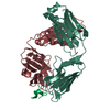 1acyS S: Starting model for refinement |
|---|---|
| Similar structure data |


-Links
 PDBj
PDBj


- Assembly
| Deposited unit | 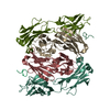
| ||||||||
|---|---|---|---|---|---|---|---|---|---|
| 1 | 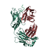
| ||||||||
| 2 | 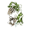
| ||||||||
| Unit cell |
|
-Components
| #1: Antibody | Mass: 24156.703 Da / Num. of mol.: 2 / Source method: isolated from a natural source / Source: (natural) #2: Antibody | Mass: 23089.838 Da / Num. of mol.: 2 / Source method: isolated from a natural source / Source: (natural) #3: Protein/peptide | Mass: 1827.182 Da / Num. of mol.: 2 / Source method: obtained synthetically Details: The peptide was synthesized chemically; its sequence occurs in the MN HIV-1 viral isolate. References: UniProt: P05877*PLUS #4: Water | ChemComp-HOH / |  Mass: 18.015 Da / Num. of mol.: 107 / Source method: isolated from a natural source / Formula: H2O Mass: 18.015 Da / Num. of mol.: 107 / Source method: isolated from a natural source / Formula: H2OHas protein modification | Y | |
|---|
-Experimental details
-Experiment
| Experiment | Method: X-RAY DIFFRACTION / Number of used crystals: 1 |
|---|
- Sample preparation
| Crystal | Density Matthews: 2.54 Å3/Da / Density % sol: 51.11 % | ||||||||||||||||||||||||
|---|---|---|---|---|---|---|---|---|---|---|---|---|---|---|---|---|---|---|---|---|---|---|---|---|---|
| Crystal grow | Temperature: 298 K / Method: vapor diffusion, sitting drop / pH: 6 Details: 1.6M Na/K phosphate, 5% isopropanol, pH 6.0, VAPOR DIFFUSION, SITTING DROP, temperature 298.K | ||||||||||||||||||||||||
| Crystal grow | *PLUS Method: vapor diffusion, sitting drop | ||||||||||||||||||||||||
| Components of the solutions | *PLUS
|
-Data collection
| Diffraction | Mean temperature: 100 K |
|---|---|
| Diffraction source | Source: SYNCHROTRON / Site: APS  / Beamline: 19-ID / Wavelength: 1.033 Å / Beamline: 19-ID / Wavelength: 1.033 Å |
| Detector | Type: CUSTOM-MADE / Detector: CCD / Date: Jun 16, 2000 |
| Radiation | Protocol: SINGLE WAVELENGTH / Monochromatic (M) / Laue (L): M / Scattering type: x-ray |
| Radiation wavelength | Wavelength: 1.033 Å / Relative weight: 1 |
| Reflection | Resolution: 2.57→48.2 Å / Num. all: 29751 / Num. obs: 29751 / % possible obs: 96.5 % / Observed criterion σ(F): 0 / Observed criterion σ(I): -3 / Redundancy: 2.9 % / Biso Wilson estimate: 42.1 Å2 / Rsym value: 0.115 / Net I/σ(I): 9.4 |
| Reflection shell | Resolution: 2.57→2.66 Å / Mean I/σ(I) obs: 2.1 / Rsym value: 0.394 / % possible all: 80 |
| Reflection | *PLUS Num. measured all: 87725 / Rmerge(I) obs: 0.115 |
| Reflection shell | *PLUS % possible obs: 80 % / Rmerge(I) obs: 0.394 |
- Processing
| Software |
| |||||||||||||||||||||||||
|---|---|---|---|---|---|---|---|---|---|---|---|---|---|---|---|---|---|---|---|---|---|---|---|---|---|---|
| Refinement | Method to determine structure: MOLECULAR REPLACEMENT Starting model: PDB ENTRY 1ACY Resolution: 2.57→48.08 Å / Rfactor Rfree error: 0.009 / Isotropic thermal model: RESTRAINED / Cross valid method: THROUGHOUT / σ(F): 0 / Stereochemistry target values: Engh & Huber Details: RESIDUES 128-135 OF CHAINS H AND I HAVE VERY WEAK ELECTRON DENSITY AND WERE GIVEN OCCUPANCY VALUES OF 0.0
| |||||||||||||||||||||||||
| Solvent computation | Solvent model: FLAT MODEL / Bsol: 38.5752 Å2 / ksol: 0.376389 e/Å3 | |||||||||||||||||||||||||
| Displacement parameters | Biso mean: 29.3 Å2
| |||||||||||||||||||||||||
| Refine analyze | Luzzati coordinate error free: 0.54 Å / Luzzati sigma a free: 0.55 Å | |||||||||||||||||||||||||
| Refinement step | Cycle: LAST / Resolution: 2.57→48.08 Å
| |||||||||||||||||||||||||
| Refine LS restraints |
| |||||||||||||||||||||||||
| LS refinement shell | Resolution: 2.57→2.66 Å / Rfactor Rfree error: 0.066 / Total num. of bins used: 20
| |||||||||||||||||||||||||
| Xplor file |
| |||||||||||||||||||||||||
| Refinement | *PLUS Lowest resolution: 48.2 Å / % reflection Rfree: 5 % | |||||||||||||||||||||||||
| Solvent computation | *PLUS | |||||||||||||||||||||||||
| Displacement parameters | *PLUS | |||||||||||||||||||||||||
| Refine LS restraints | *PLUS
|
