 Movie
Movie Controller

[English] 日本語
 Yorodumi
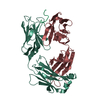
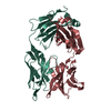
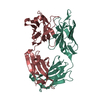
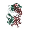
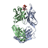
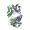
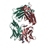
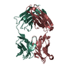
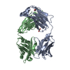
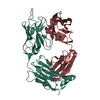
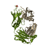
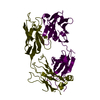
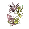
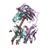
- PDB-4g6k: Crystal structure of the therapeutic antibody binding fragment of... -
+ Open data

- Basic information
| Entry | Database: PDB / ID: 4g6k | ||||||
|---|---|---|---|---|---|---|---|
| Title | Crystal structure of the therapeutic antibody binding fragment of gevokizumab in its unbound state | ||||||
 Components Components |
| ||||||
 Keywords Keywords | IMMUNE SYSTEM / immunoglobulin fold / cytokine / interleukine-1beta | ||||||
| Function / homology | Immunoglobulins / Immunoglobulin-like / Sandwich / Mainly Beta Function and homology information Function and homology information | ||||||
| Biological species |  homo Sapiens (human) homo Sapiens (human) | ||||||
| Method |  X-RAY DIFFRACTION / SYNCHROTRON / MOLECULAR REPLACEMENT / Resolution: 1.9 Å X-RAY DIFFRACTION / SYNCHROTRON / MOLECULAR REPLACEMENT / Resolution: 1.9 Å | ||||||
 Authors Authors | Blech, M. / Hoerer, S. | ||||||
 Citation Citation | Journal: J.Mol.Biol. / Year: 2013 Title: One target-two different binding modes: Structural insights into gevokizumab and canakinumab interactions to interleukin-1beta Authors: Blech, M. / Peter, D. / Fischer, P. / Bauer, M.M. / Hafner, M. / Zeeb, M. / Nar, H. | ||||||
| History |
|
- Structure visualization
| Structure viewer | Molecule: MolmilJmol/JSmol |
|---|
- Downloads & links
-Download
| PDBx/mmCIF format | 4g6k.cif.gz | 185.4 KB | Display | PDBx/mmCIF format |
|---|---|---|---|---|
| PDB format | pdb4g6k.ent.gz | 147.9 KB | Display | PDB format |
| PDBx/mmJSON format | 4g6k.json.gz | Tree view | PDBx/mmJSON format | |
| Others |  Other downloads Other downloads |
-Validation report
| Arichive directory | https://data.pdbj.org/pub/pdb/validation_reports/g6/4g6kftp://data.pdbj.org/pub/pdb/validation_reports/g6/4g6k | HTTPS FTP |
|---|
-Related structure data
| Related structure data |  4g5zC  4g6jC  4g6mC 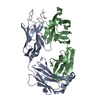 1pz5S  3bkjS C: citing same article ( S: Starting model for refinement |
|---|---|
| Similar structure data |


-Links
 PDBj
PDBj

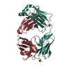
- Assembly
| Deposited unit | 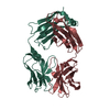
| ||||||||
|---|---|---|---|---|---|---|---|---|---|
| 1 |
| ||||||||
| Unit cell |
|
-Components
| #1: Antibody | Mass: 23420.236 Da / Num. of mol.: 1 Source method: isolated from a genetically manipulated source Source: (gene. exp.) homo Sapiens, Mus musculus / Cell line (production host): CHO / Production host:  Cricetulus griseus (Chinese hamster) Cricetulus griseus (Chinese hamster) |
|---|---|
| #2: Antibody | Mass: 23371.086 Da / Num. of mol.: 1 Source method: isolated from a genetically manipulated source Source: (gene. exp.) homo Sapiens, Mus musculus / Cell line (production host): CHO / Production host: Cricetulus griseus (Chinese hamster) |
| #3: Water | ChemComp-HOH /  Mass: 18.015 Da / Num. of mol.: 510 / Source method: isolated from a natural source / Formula: H2O Mass: 18.015 Da / Num. of mol.: 510 / Source method: isolated from a natural source / Formula: H2O |
| Has protein modification | Y |
-Experimental details
-Experiment
| Experiment | Method: X-RAY DIFFRACTION / Number of used crystals: 1 |
|---|
- Sample preparation
| Crystal | Density Matthews: 2.65 Å3/Da / Density % sol: 53.59 % |
|---|---|
| Crystal grow | Temperature: 293 K / Method: vapor diffusion, sitting drop / pH: 5 Details: 14.3% w/v PEG 3350 and 0.14M tri-sodium citrate at pH 5.0 equilibrated against 24% w/v PEG 3350, VAPOR DIFFUSION, SITTING DROP, temperature 293K |
-Data collection
| Diffraction | Mean temperature: 100 K |
|---|---|
| Diffraction source | Source: SYNCHROTRON / Site: SLS  / Beamline: X06SA / Wavelength: 0.91 Å / Beamline: X06SA / Wavelength: 0.91 Å |
| Detector | Type: PSI PILATUS 6M / Detector: PIXEL / Date: Nov 22, 2011 |
| Radiation | Protocol: SINGLE WAVELENGTH / Monochromatic (M) / Laue (L): M / Scattering type: x-ray |
| Radiation wavelength | Wavelength: 0.91 Å / Relative weight: 1 |
| Reflection | Resolution: 1.9→57.4 Å / Num. all: 284279 / Num. obs: 41364 / % possible obs: 100 % / Observed criterion σ(F): 2 / Observed criterion σ(I): 2 / Redundancy: 6.5 % / Biso Wilson estimate: 24.43 Å2 / Rmerge(I) obs: 0.076 / Rsym value: 0.084 / Net I/σ(I): 19.4 |
| Reflection shell | Highest resolution: 1.9 Å |
- Processing
| Software |
| ||||||||||||||||||||||||||||||||||||||||||||||||||||||||||||||||||||||||||||||||||||||||||||||||||||||||||||||||||
|---|---|---|---|---|---|---|---|---|---|---|---|---|---|---|---|---|---|---|---|---|---|---|---|---|---|---|---|---|---|---|---|---|---|---|---|---|---|---|---|---|---|---|---|---|---|---|---|---|---|---|---|---|---|---|---|---|---|---|---|---|---|---|---|---|---|---|---|---|---|---|---|---|---|---|---|---|---|---|---|---|---|---|---|---|---|---|---|---|---|---|---|---|---|---|---|---|---|---|---|---|---|---|---|---|---|---|---|---|---|---|---|---|---|---|---|
| Refinement | Method to determine structure: MOLECULAR REPLACEMENT Starting model: 1PZ5 and 3BKJ Resolution: 1.9→27.75 Å / Cor.coef. Fo:Fc: 0.9458 / Cor.coef. Fo:Fc free: 0.9335 / SU R Cruickshank DPI: 0.136 / Cross valid method: THROUGHOUT / σ(F): 0
| ||||||||||||||||||||||||||||||||||||||||||||||||||||||||||||||||||||||||||||||||||||||||||||||||||||||||||||||||||
| Displacement parameters | Biso mean: 28.32 Å2
| ||||||||||||||||||||||||||||||||||||||||||||||||||||||||||||||||||||||||||||||||||||||||||||||||||||||||||||||||||
| Refine analyze | Luzzati coordinate error obs: 0.212 Å | ||||||||||||||||||||||||||||||||||||||||||||||||||||||||||||||||||||||||||||||||||||||||||||||||||||||||||||||||||
| Refinement step | Cycle: LAST / Resolution: 1.9→27.75 Å
| ||||||||||||||||||||||||||||||||||||||||||||||||||||||||||||||||||||||||||||||||||||||||||||||||||||||||||||||||||
| Refine LS restraints |
| ||||||||||||||||||||||||||||||||||||||||||||||||||||||||||||||||||||||||||||||||||||||||||||||||||||||||||||||||||
| LS refinement shell | Resolution: 1.9→1.95 Å / Total num. of bins used: 20
| ||||||||||||||||||||||||||||||||||||||||||||||||||||||||||||||||||||||||||||||||||||||||||||||||||||||||||||||||||
| Refinement TLS params. | Method: refined / Refine-ID: X-RAY DIFFRACTION
| ||||||||||||||||||||||||||||||||||||||||||||||||||||||||||||||||||||||||||||||||||||||||||||||||||||||||||||||||||
| Refinement TLS group |
|
