 Movie
Movie Controller

[English] 日本語
 Yorodumi
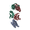
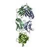
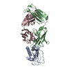
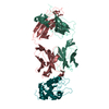
- PDB-4g6j: Crystal structure of human IL-1beta in complex with the therapeut... -
+ Open data

- Basic information
| Entry | Database: PDB / ID: 4g6j | ||||||
|---|---|---|---|---|---|---|---|
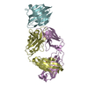
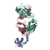
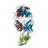
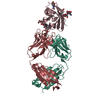
| Title | Crystal structure of human IL-1beta in complex with the therapeutic antibody binding fragment of canakinumab | ||||||
 Components Components |
| ||||||
 Keywords Keywords | IMMUNE SYSTEM / immunglobulin fold / interleukin-1beta binding | ||||||
| Function / homology |  Function and homology information Function and homology informationpositive regulation of T cell mediated immunity / negative regulation of adiponectin secretion / negative regulation of lipid metabolic process / smooth muscle adaptation / positive regulation of lipid catabolic process / positive regulation of cell adhesion molecule production / regulation of nitric-oxide synthase activity / negative regulation of D-glucose transmembrane transport / positive regulation of T-helper 1 cell cytokine production / hyaluronan biosynthetic process ...positive regulation of T cell mediated immunity / negative regulation of adiponectin secretion / negative regulation of lipid metabolic process / smooth muscle adaptation / positive regulation of lipid catabolic process / positive regulation of cell adhesion molecule production / regulation of nitric-oxide synthase activity / negative regulation of D-glucose transmembrane transport / positive regulation of T-helper 1 cell cytokine production / hyaluronan biosynthetic process / positive regulation of complement activation / positive regulation of RNA biosynthetic process / cellular response to interleukin-17 / monocyte aggregation / positive regulation of tight junction disassembly / negative regulation of gap junction assembly / positive regulation of prostaglandin secretion / positive regulation of immature T cell proliferation in thymus / vascular endothelial growth factor production / positive regulation of prostaglandin biosynthetic process / positive regulation of fever generation / positive regulation of platelet-derived growth factor receptor signaling pathway / regulation of defense response to virus by host / fever generation / regulation of establishment of endothelial barrier / CLEC7A/inflammasome pathway / Interleukin-1 processing / positive regulation of macrophage derived foam cell differentiation / positive regulation of monocyte chemotactic protein-1 production / interleukin-1 receptor binding / positive regulation of p38MAPK cascade / negative regulation of synaptic transmission / response to carbohydrate / positive regulation of vascular endothelial growth factor receptor signaling pathway / positive regulation of granulocyte macrophage colony-stimulating factor production / positive regulation of heterotypic cell-cell adhesion / positive regulation of membrane protein ectodomain proteolysis / regulation of canonical NF-kappaB signal transduction / interleukin-1-mediated signaling pathway / positive regulation of neuroinflammatory response / Interleukin-10 signaling / response to ATP / regulation of insulin secretion / positive regulation of cell division / positive regulation of vascular endothelial growth factor production / positive regulation of glial cell proliferation / Pyroptosis / regulation of neurogenesis / negative regulation of extrinsic apoptotic signaling pathway in absence of ligand / ectopic germ cell programmed cell death / positive regulation of epithelial to mesenchymal transition / negative regulation of lipid catabolic process / regulation of ERK1 and ERK2 cascade / positive regulation of interleukin-2 production / Purinergic signaling in leishmaniasis infection / JNK cascade / extrinsic apoptotic signaling pathway in absence of ligand / negative regulation of MAPK cascade / neutrophil chemotaxis / positive regulation of mitotic nuclear division / negative regulation of insulin receptor signaling pathway / embryo implantation / positive regulation of T cell proliferation / secretory granule / astrocyte activation / response to interleukin-1 / positive regulation of protein export from nucleus / cytokine activity / positive regulation of interleukin-8 production / cellular response to mechanical stimulus / positive regulation of non-canonical NF-kappaB signal transduction / negative regulation of neurogenesis / positive regulation of interleukin-6 production / positive regulation of JNK cascade / integrin binding / cellular response to xenobiotic stimulus / positive regulation of type II interferon production / cytokine-mediated signaling pathway / positive regulation of angiogenesis / Interleukin-1 signaling / positive regulation of nitric oxide biosynthetic process / positive regulation of inflammatory response / cell-cell signaling / cellular response to lipopolysaccharide / Interleukin-4 and Interleukin-13 signaling / response to lipopolysaccharide / positive regulation of canonical NF-kappaB signal transduction / positive regulation of MAPK cascade / positive regulation of ERK1 and ERK2 cascade / lysosome / positive regulation of phosphatidylinositol 3-kinase/protein kinase B signal transduction / defense response to Gram-positive bacterium / immune response / positive regulation of cell migration / inflammatory response / negative regulation of cell population proliferation / protein domain specific binding / apoptotic process / positive regulation of cell population proliferation / positive regulation of gene expression Similarity search - Function | ||||||
| Biological species |  Homo sapiens (human) Homo sapiens (human) | ||||||
| Method |  X-RAY DIFFRACTION / SYNCHROTRON / MOLECULAR REPLACEMENT / Resolution: 2.03 Å X-RAY DIFFRACTION / SYNCHROTRON / MOLECULAR REPLACEMENT / Resolution: 2.03 Å | ||||||
 Authors Authors | Blech, M. / Hoerer, S. | ||||||
 Citation Citation | Journal: J.Mol.Biol. / Year: 2013 Title: One traget-two different binding modes: Structural insights into gevokizumab and canakinumab interactions to interleukin-1beta Authors: Blech, M. / Peter, D. / Fischer, P. / Bauer, M.M. / Hafner, M. / Zeeb, M. / Nar, H. | ||||||
| History |
|
- Structure visualization
| Structure viewer | Molecule: MolmilJmol/JSmol |
|---|
- Downloads & links
-Download
| PDBx/mmCIF format | 4g6j.cif.gz | 240.7 KB | Display | PDBx/mmCIF format |
|---|---|---|---|---|
| PDB format | pdb4g6j.ent.gz | 194.2 KB | Display | PDB format |
| PDBx/mmJSON format | 4g6j.json.gz | Tree view | PDBx/mmJSON format | |
| Others |  Other downloads Other downloads |
-Validation report
| Arichive directory | https://data.pdbj.org/pub/pdb/validation_reports/g6/4g6jftp://data.pdbj.org/pub/pdb/validation_reports/g6/4g6j | HTTPS FTP |
|---|
-Related structure data
| Related structure data |  4g5zC  4g6kC  4g6mC 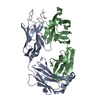 1pz5S  1tooS  3bkjS C: citing same article ( S: Starting model for refinement |
|---|---|
| Similar structure data |

-Links
 PDBj
PDBj


- Assembly
| Deposited unit | 
| ||||||||
|---|---|---|---|---|---|---|---|---|---|
| 1 |
| ||||||||
| Unit cell |
|
-Components
| #1: Protein | Mass: 17993.629 Da / Num. of mol.: 1 / Fragment: human interleukin-1beta / Source method: obtained synthetically / Source: (synth.) Homo sapiens (human) / References: UniProt: P01584 |
|---|---|
| #2: Antibody | Mass: 23349.152 Da / Num. of mol.: 1 Fragment: heavy chain of antibody binding fragment of canakinumab Source method: obtained synthetically / Source: (synth.) Homo sapiens (human) |
| #3: Antibody | Mass: 23149.652 Da / Num. of mol.: 1 Fragment: light chain of antibody binding fragment of canakinumab Source method: obtained synthetically / Source: (synth.) Homo sapiens (human) |
| #4: Water | ChemComp-HOH /  Mass: 18.015 Da / Num. of mol.: 303 / Source method: isolated from a natural source / Formula: H2O Mass: 18.015 Da / Num. of mol.: 303 / Source method: isolated from a natural source / Formula: H2O |
| Has protein modification | Y |
-Experimental details
-Experiment
| Experiment | Method: X-RAY DIFFRACTION / Number of used crystals: 1 |
|---|
- Sample preparation
| Crystal | Density Matthews: 2.22 Å3/Da / Density % sol: 44.6 % |
|---|---|
| Crystal grow | Temperature: 293 K / Method: vapor diffusion, sitting drop / pH: 4.5 Details: 17.2% w/v PEG 3350 and 0.14M tri-sodium citrate, 24% w/v PEG 3350, pH 4.5, VAPOR DIFFUSION, SITTING DROP, temperature 293K |
-Data collection
| Diffraction | Mean temperature: 100 K |
|---|---|
| Diffraction source | Source: SYNCHROTRON / Site: SLS  / Beamline: X06SA / Wavelength: 0.91 Å / Beamline: X06SA / Wavelength: 0.91 Å |
| Detector | Type: PSI PILATUS 6M / Detector: PIXEL / Date: Mar 9, 2012 |
| Radiation | Protocol: SINGLE WAVELENGTH / Monochromatic (M) / Laue (L): M / Scattering type: x-ray |
| Radiation wavelength | Wavelength: 0.91 Å / Relative weight: 1 |
| Reflection | Resolution: 2→94.7 Å / Num. all: 207933 / Num. obs: 38275 / % possible obs: 98.8 % / Observed criterion σ(F): 2 / Observed criterion σ(I): 2 / Redundancy: 5.3 % / Biso Wilson estimate: 29.23 Å2 / Rmerge(I) obs: 0.108 / Rsym value: 0.12 / Net I/σ(I): 12.1 |
| Reflection shell | Resolution: 2→2.1 Å / Redundancy: 5.3 % / Rmerge(I) obs: 0.108 / Mean I/σ(I) obs: 2.3 / Num. unique all: 37806 / Rsym value: 0.12 / % possible all: 93.5 |
- Processing
| Software |
| ||||||||||||||||||||||||||||||||||||||||||||||||||||||||||||||||||||||||||||||||||||||||||||||||||||
|---|---|---|---|---|---|---|---|---|---|---|---|---|---|---|---|---|---|---|---|---|---|---|---|---|---|---|---|---|---|---|---|---|---|---|---|---|---|---|---|---|---|---|---|---|---|---|---|---|---|---|---|---|---|---|---|---|---|---|---|---|---|---|---|---|---|---|---|---|---|---|---|---|---|---|---|---|---|---|---|---|---|---|---|---|---|---|---|---|---|---|---|---|---|---|---|---|---|---|---|---|---|
| Refinement | Method to determine structure: MOLECULAR REPLACEMENT Starting model: 1PZ5, 3BKJ, 1TOO Resolution: 2.03→39.36 Å / Cor.coef. Fo:Fc: 0.9065 / Cor.coef. Fo:Fc free: 0.8926 / SU R Cruickshank DPI: 0.231 / Cross valid method: THROUGHOUT / σ(F): 0 / σ(I): 2
| ||||||||||||||||||||||||||||||||||||||||||||||||||||||||||||||||||||||||||||||||||||||||||||||||||||
| Displacement parameters | Biso mean: 35.92 Å2
| ||||||||||||||||||||||||||||||||||||||||||||||||||||||||||||||||||||||||||||||||||||||||||||||||||||
| Refine analyze | Luzzati coordinate error obs: 0.3 Å | ||||||||||||||||||||||||||||||||||||||||||||||||||||||||||||||||||||||||||||||||||||||||||||||||||||
| Refinement step | Cycle: LAST / Resolution: 2.03→39.36 Å
| ||||||||||||||||||||||||||||||||||||||||||||||||||||||||||||||||||||||||||||||||||||||||||||||||||||
| Refine LS restraints |
| ||||||||||||||||||||||||||||||||||||||||||||||||||||||||||||||||||||||||||||||||||||||||||||||||||||
| LS refinement shell | Resolution: 2.03→2.09 Å / Total num. of bins used: 19
| ||||||||||||||||||||||||||||||||||||||||||||||||||||||||||||||||||||||||||||||||||||||||||||||||||||
| Refinement TLS params. | Method: refined / Refine-ID: X-RAY DIFFRACTION
| ||||||||||||||||||||||||||||||||||||||||||||||||||||||||||||||||||||||||||||||||||||||||||||||||||||
| Refinement TLS group |
|
