 Movie
Movie Controller

[English] 日本語
 Yorodumi
- PDB-4yc2: Crystal structure of the stabilized inner domain of clade A/E HIV... -
+ Open data

- Basic information
| Entry | Database: PDB / ID: 4yc2 | ||||||
|---|---|---|---|---|---|---|---|
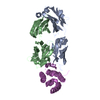
| Title | Crystal structure of the stabilized inner domain of clade A/E HIV-1 gp120 from E. coli in complex with the antibody A32. | ||||||
 Components Components |
| ||||||
 Keywords Keywords | VIRAL PROTEIN/IMMUNE SYSTEM / ADCC / NON-NEUTRALIZING / ANTI-HIV-1 ENV ANTIBODY A32 / CD4I ANTIBODY / CLADE A/E 93TH057 / VIRAL PROTEIN / VIRAL PROTEIN-IMMUNE SYSTEM complex | ||||||
| Function / homology |  Function and homology information Function and homology informationmembrane fusion involved in viral entry into host cell / viral envelope / symbiont entry into host cell / virion attachment to host cell / virion membrane Similarity search - Function | ||||||
| Biological species |   Human immunodeficiency virus 1 Human immunodeficiency virus 1 Homo sapiens (human) Homo sapiens (human) | ||||||
| Method |  X-RAY DIFFRACTION / SYNCHROTRON / MOLECULAR REPLACEMENT / Resolution: 3.02 Å X-RAY DIFFRACTION / SYNCHROTRON / MOLECULAR REPLACEMENT / Resolution: 3.02 Å | ||||||
 Authors Authors | Tolbert, W.D. / Gohain, N. / Pazgier, M. | ||||||
| Funding support |  United States, 1items United States, 1items
| ||||||
 Citation Citation | Journal: Structure / Year: 2016 Title: Paring Down HIV Env: Design and Crystal Structure of a Stabilized Inner Domain of HIV-1 gp120 Displaying a Major ADCC Target of the A32 Region. Authors: Tolbert, W.D. / Gohain, N. / Veillette, M. / Chapleau, J.P. / Orlandi, C. / Visciano, M.L. / Ebadi, M. / DeVico, A.L. / Fouts, T.R. / Finzi, A. / Lewis, G.K. / Pazgier, M. | ||||||
| History |
|
- Structure visualization
| Structure viewer | Molecule: MolmilJmol/JSmol |
|---|
- Downloads & links
-Download
| PDBx/mmCIF format | 4yc2.cif.gz | 438.6 KB | Display | PDBx/mmCIF format |
|---|---|---|---|---|
| PDB format | pdb4yc2.ent.gz | 364 KB | Display | PDB format |
| PDBx/mmJSON format | 4yc2.json.gz | Tree view | PDBx/mmJSON format | |
| Others |  Other downloads Other downloads |
-Validation report
| Arichive directory | https://data.pdbj.org/pub/pdb/validation_reports/yc/4yc2ftp://data.pdbj.org/pub/pdb/validation_reports/yc/4yc2 | HTTPS FTP |
|---|
-Related structure data
| Related structure data | 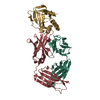 4yblC 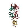 5fcuC 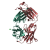 3tnmS  4rqh S: Starting model for refinement C: citing same article ( |
|---|---|
| Similar structure data |


-Links
 PDBj
PDBj


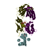
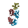
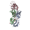
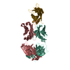
- Assembly
| Deposited unit | 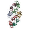
| ||||||||
|---|---|---|---|---|---|---|---|---|---|
| 1 | 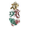
| ||||||||
| 2 | 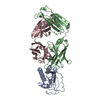
| ||||||||
| Unit cell |
| ||||||||
| Details | Complexes were purified by gel filtration prior to crystallization |
-Components
| #1: Protein | Mass: 17380.535 Da / Num. of mol.: 2 / Mutation: V65C, S115C Source method: isolated from a genetically manipulated source Source: (gene. exp.) Human immunodeficiency virus 1 / Variant: clade A/E / Production host:  #2: Antibody | Mass: 23991.855 Da / Num. of mol.: 2 Source method: isolated from a genetically manipulated source Source: (gene. exp.) Homo sapiens (human) / Cell line (production host): HEK 293 cells / Production host: Homo sapiens (human)#3: Antibody | Mass: 22193.434 Da / Num. of mol.: 2 Source method: isolated from a genetically manipulated source Source: (gene. exp.) Homo sapiens (human) / Cell line (production host): HEK 293 cells / Production host: Homo sapiens (human)#4: Water | ChemComp-HOH / |  Mass: 18.015 Da / Num. of mol.: 4 / Source method: isolated from a natural source / Formula: H2O Mass: 18.015 Da / Num. of mol.: 4 / Source method: isolated from a natural source / Formula: H2OHas protein modification | Y | Sequence details | The sequence of the clade A/E gp120 is based on the HIV-1 clade A/E gp120 sequence in PDB ID 3TGT. ...The sequence of the clade A/E gp120 is based on the HIV-1 clade A/E gp120 sequence in PDB ID 3TGT. The sequence was engineered to remove the outer domain of gp120 and consists of the N-terminal sequence plus some of the C-terminal sequence. The numbering in the PDB file is consistent with 3TGT and 4H8W and is based on the Hxbc2 gp120 sequence which serves as the reference sequence for most gp120 structures. | |
|---|
-Experimental details
-Experiment
| Experiment | Method: X-RAY DIFFRACTION |
|---|
- Sample preparation
| Crystal | Density Matthews: 2.18 Å3/Da / Density % sol: 43.7 % |
|---|---|
| Crystal grow | Temperature: 295 K / Method: vapor diffusion, hanging drop / pH: 8.5 / Details: 18-22% PEG 6000 or PEG8000 0.1 M Tris-HCl pH 8.5 |
-Data collection
| Diffraction | Mean temperature: 100 K |
|---|---|
| Diffraction source | Source: SYNCHROTRON / Site: SSRL / Beamline: BL12-2 / Wavelength: 0.97946 Å |
| Detector | Type: PSI PILATUS 6M / Detector: PIXEL / Date: Jan 23, 2015 |
| Radiation | Monochromator: Si(111) / Protocol: SINGLE WAVELENGTH / Monochromatic (M) / Laue (L): M / Scattering type: x-ray |
| Radiation wavelength | Wavelength: 0.97946 Å / Relative weight: 1 |
| Reflection | Resolution: 3→50 Å / Num. all: 23714 / Num. obs: 20655 / % possible obs: 87.1 % / Observed criterion σ(F): 0 / Observed criterion σ(I): 0 / Redundancy: 5.8 % / Rmerge(I) obs: 0.163 / Net I/σ(I): 9.5 |
| Reflection shell | Resolution: 3→3.05 Å / Redundancy: 5.8 % / Rmerge(I) obs: 0.88 / Mean I/σ(I) obs: 1.2 / % possible all: 86.7 |
- Processing
| Software |
| ||||||||||||||||||||||||||||||||||||||||||||||||||||||||||||||||||||||||||||||||||||||||||||||||||||||||||||||||||||||||||||||||||||||||||||||||||||||||||||||||||||||||||||||||||||||
|---|---|---|---|---|---|---|---|---|---|---|---|---|---|---|---|---|---|---|---|---|---|---|---|---|---|---|---|---|---|---|---|---|---|---|---|---|---|---|---|---|---|---|---|---|---|---|---|---|---|---|---|---|---|---|---|---|---|---|---|---|---|---|---|---|---|---|---|---|---|---|---|---|---|---|---|---|---|---|---|---|---|---|---|---|---|---|---|---|---|---|---|---|---|---|---|---|---|---|---|---|---|---|---|---|---|---|---|---|---|---|---|---|---|---|---|---|---|---|---|---|---|---|---|---|---|---|---|---|---|---|---|---|---|---|---|---|---|---|---|---|---|---|---|---|---|---|---|---|---|---|---|---|---|---|---|---|---|---|---|---|---|---|---|---|---|---|---|---|---|---|---|---|---|---|---|---|---|---|---|---|---|---|---|
| Refinement | Method to determine structure: MOLECULAR REPLACEMENT Starting model: 3TNM and 4RQH Resolution: 3.02→50 Å / Cor.coef. Fo:Fc: 0.933 / Cor.coef. Fo:Fc free: 0.888 / SU B: 72.5 / SU ML: 0.556 / Cross valid method: THROUGHOUT / ESU R Free: 0.616 / Stereochemistry target values: MAXIMUM LIKELIHOOD / Details: HYDROGENS HAVE BEEN ADDED IN THE RIDING POSITIONS
| ||||||||||||||||||||||||||||||||||||||||||||||||||||||||||||||||||||||||||||||||||||||||||||||||||||||||||||||||||||||||||||||||||||||||||||||||||||||||||||||||||||||||||||||||||||||
| Solvent computation | Ion probe radii: 0.8 Å / Shrinkage radii: 0.8 Å / VDW probe radii: 1.2 Å / Solvent model: MASK | ||||||||||||||||||||||||||||||||||||||||||||||||||||||||||||||||||||||||||||||||||||||||||||||||||||||||||||||||||||||||||||||||||||||||||||||||||||||||||||||||||||||||||||||||||||||
| Displacement parameters | Biso mean: 88.657 Å2
| ||||||||||||||||||||||||||||||||||||||||||||||||||||||||||||||||||||||||||||||||||||||||||||||||||||||||||||||||||||||||||||||||||||||||||||||||||||||||||||||||||||||||||||||||||||||
| Refinement step | Cycle: 1 / Resolution: 3.02→50 Å
| ||||||||||||||||||||||||||||||||||||||||||||||||||||||||||||||||||||||||||||||||||||||||||||||||||||||||||||||||||||||||||||||||||||||||||||||||||||||||||||||||||||||||||||||||||||||
| Refine LS restraints |
|