 Movie
Movie Controller


+ Open data

- Basic information
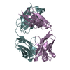
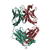
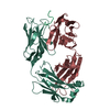
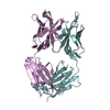
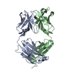
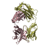
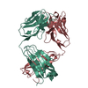
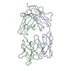
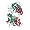
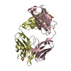
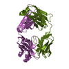
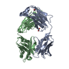
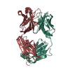
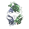
| Entry | Database: PDB / ID: 1zan | ||||||
|---|---|---|---|---|---|---|---|
| Title | Crystal structure of anti-NGF AD11 Fab | ||||||
 Components Components |
| ||||||
 Keywords Keywords | IMMUNE SYSTEM / Immunoglobulin / Fab | ||||||
| Function / homology |  Function and homology information Function and homology informationimmunoglobulin mediated immune response / immunoglobulin complex / antigen binding / adaptive immune response / extracellular region / plasma membrane Similarity search - Function | ||||||
| Biological species |  | ||||||
| Method |  X-RAY DIFFRACTION / SYNCHROTRON / MOLECULAR REPLACEMENT / Resolution: 1.7 Å X-RAY DIFFRACTION / SYNCHROTRON / MOLECULAR REPLACEMENT / Resolution: 1.7 Å | ||||||
 Authors Authors | Covaceuszach, S. / Cattaneo, A. / Cassetta, A. / Lamba, D. | ||||||
 Citation Citation | Journal: J Mol Biol / Year: 2008 Title: Dissecting NGF interactions with TrkA and p75 receptors by structural and functional studies of an anti-NGF neutralizing antibody. Authors: Sonia Covaceuszach / Alberto Cassetta / Petr V Konarev / Stefania Gonfloni / Rainer Rudolph / Dmitri I Svergun / Doriano Lamba / Antonino Cattaneo /  Abstract: The anti-nerve growth factor (NGF) monoclonal antibody alphaD11 is a potent antagonist that neutralizes the biological functions of its antigen in vivo. NGF antagonism is expected to be a highly ...The anti-nerve growth factor (NGF) monoclonal antibody alphaD11 is a potent antagonist that neutralizes the biological functions of its antigen in vivo. NGF antagonism is expected to be a highly effective and safe therapeutic approach in many pain states. A comprehensive functional and structural analysis of alphaD11 monoclonal antibody was carried out, showing its ability to neutralize NGF binding to either tropomyosine receptor kinase A (TrkA) or p75 receptors. The 3-D structure of the alphaD11 Fab fragment was solved at 1.7 A resolution. A computational docking model of the alphaD11 Fab-NGF complex, based on epitope mapping using a pool of 44 NGF mutants and experimentally validated by small-angle X-ray scattering, provided the structural basis for identifying the residues involved in alphaD11 Fab binding. The present study pinpoints loop II of NGF to be an important structural determinant for NGF biological activity mediated by TrkA receptor. #1: Journal: ACTA CRYSTALLOGR.,SECT.D / Year: 2004Title: Purification, crystallization, X-ray diffraction analysis and phasing of a Fab fragment of monoclonal neuroantibody AD11 against nerve growth factor. Authors: Covaceuszach, S. / Cassetta, A. / Cattaneo, A. / Lamba, D. | ||||||
| History |
|
- Structure visualization
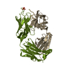
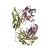
| Structure viewer | Molecule: MolmilJmol/JSmol |
|---|
- Downloads & links
-Download
| PDBx/mmCIF format | 1zan.cif.gz | 104.2 KB | Display | PDBx/mmCIF format |
|---|---|---|---|---|
| PDB format | pdb1zan.ent.gz | 77.7 KB | Display | PDB format |
| PDBx/mmJSON format | 1zan.json.gz | Tree view | PDBx/mmJSON format | |
| Others |  Other downloads Other downloads |
-Validation report
| Arichive directory | https://data.pdbj.org/pub/pdb/validation_reports/za/1zanftp://data.pdbj.org/pub/pdb/validation_reports/za/1zan | HTTPS FTP |
|---|
-Related structure data
| Related structure data |  1cicS S: Starting model for refinement C: citing same article ( |
|---|---|
| Similar structure data |


-Links
 PDBj
PDBj

- Assembly
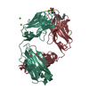
| Deposited unit | 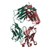
| ||||||||
|---|---|---|---|---|---|---|---|---|---|
| 1 |
| ||||||||
| Unit cell |
|
-Components
| #1: Antibody | Mass: 23340.736 Da / Num. of mol.: 1 Source method: isolated from a genetically manipulated source Source: (gene. exp.) Keywords: Light Chain / References: UniProt: Q4KM66 |
|---|---|
| #2: Antibody | Mass: 23549.291 Da / Num. of mol.: 1 Source method: isolated from a genetically manipulated source Source: (gene. exp.) Keywords: Heavy Chain / References: UniProt: P84751 |
| #3: Chemical | ChemComp-CL /   Mass: 35.453 Da / Num. of mol.: 1 / Source method: obtained synthetically / Formula: Cl Mass: 35.453 Da / Num. of mol.: 1 / Source method: obtained synthetically / Formula: Cl |
| #4: Water | ChemComp-HOH /  Mass: 18.015 Da / Num. of mol.: 404 / Source method: isolated from a natural source / Formula: H2O Mass: 18.015 Da / Num. of mol.: 404 / Source method: isolated from a natural source / Formula: H2O |
| Has protein modification | Y |
-Experimental details
-Experiment
| Experiment | Method: X-RAY DIFFRACTION / Number of used crystals: 1 |
|---|
- Sample preparation
| Crystal | Density Matthews: 2.4 Å3/Da / Density % sol: 48 % |
|---|---|
| Crystal grow | Temperature: 298 K / Method: vapor diffusion, hanging drop / pH: 5.5 Details: 20% Peg4000, 600mM NaCl, 100mM BTP, pH 5.5, VAPOR DIFFUSION, HANGING DROP, temperature 298K |
-Data collection
| Diffraction | Mean temperature: 100 K |
|---|---|
| Diffraction source | Source: SYNCHROTRON / Site: ESRF  / Beamline: ID14-1 / Wavelength: 0.934 / Wavelength: 0.934 Å / Beamline: ID14-1 / Wavelength: 0.934 / Wavelength: 0.934 Å |
| Detector | Type: MARRESEARCH / Detector: CCD / Date: Nov 26, 2001 / Details: Sagitally focusing Ge(220) and a multilayer |
| Radiation | Monochromator: Diamond (111), Ge(220) / Protocol: SINGLE WAVELENGTH / Monochromatic (M) / Laue (L): M / Scattering type: x-ray |
| Radiation wavelength | Wavelength: 0.934 Å / Relative weight: 1 |
| Reflection | Resolution: 1.7→29.64 Å / Num. all: 47951 / Num. obs: 47951 / % possible obs: 97.2 % / Observed criterion σ(F): -3 / Observed criterion σ(I): 0 / Redundancy: 6.1 % / Biso Wilson estimate: 18.9 Å2 / Rsym value: 0.058 / Net I/σ(I): 5.8 |
| Reflection shell | Resolution: 1.7→1.75 Å / Redundancy: 3.7 % / Mean I/σ(I) obs: 5.5 / Num. unique all: 3198 / Rsym value: 0.278 / % possible all: 78.4 |
- Processing
| Software |
| |||||||||||||||||||||||||
|---|---|---|---|---|---|---|---|---|---|---|---|---|---|---|---|---|---|---|---|---|---|---|---|---|---|---|
| Refinement | Method to determine structure: MOLECULAR REPLACEMENT Starting model: PDB ENTRY 1CIC Resolution: 1.7→17 Å / Isotropic thermal model: Restrained / Cross valid method: THROUGHOUT / σ(F): 0 / Stereochemistry target values: Engh & Huber
| |||||||||||||||||||||||||
| Displacement parameters | Biso mean: 27.7 Å2
| |||||||||||||||||||||||||
| Refine analyze |
| |||||||||||||||||||||||||
| Refinement step | Cycle: LAST / Resolution: 1.7→17 Å
| |||||||||||||||||||||||||
| Refine LS restraints |
| |||||||||||||||||||||||||
| LS refinement shell | Resolution: 1.7→1.81 Å / Rfactor Rfree error: 0.012
|
