 Movie
Movie Controller

[English] 日本語
 Yorodumi
- PDB-2v7h: Crystal structure of an immunogen specific anti-mannopyranoside m... -
+ Open data

- Basic information
| Entry | Database: PDB / ID: 2v7h | ||||||
|---|---|---|---|---|---|---|---|
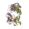
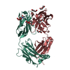
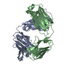
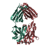
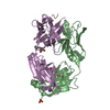
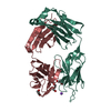
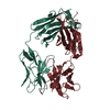
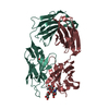
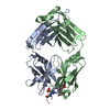
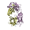
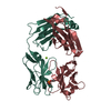
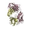
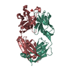
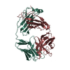
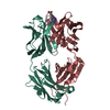
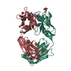
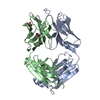
| Title | Crystal structure of an immunogen specific anti-mannopyranoside monoclonal antibody Fab fragment | ||||||
 Components Components | (MONOCLONAL ANTIBODY) x 2 | ||||||
 Keywords Keywords | IMMUNE SYSTEM / MONOCLONAL ANTIBODY / MANNOPYRANOSIDE SPECIFICITY / MOLECULAR MIMICRY | ||||||
| Function / homology | Immunoglobulins / Immunoglobulin-like / Sandwich / Mainly Beta Function and homology information Function and homology information | ||||||
| Biological species |  | ||||||
| Method |  X-RAY DIFFRACTION / MOLECULAR REPLACEMENT / Resolution: 2.8 Å X-RAY DIFFRACTION / MOLECULAR REPLACEMENT / Resolution: 2.8 Å | ||||||
 Authors Authors | Krishnan, L. / Sahni, G. / Kaur, K.J. / Salunke, D.M. | ||||||
 Citation Citation | Journal: Biophys.J. / Year: 2008 Title: Role of Antibody Paratope Conformational Flexibility in the Manifestation of Molecular Mimicry. Authors: Krishnan, L. / Sahni, G. / Kaur, K.J. / Salunke, D.M. | ||||||
| History |
| ||||||
| Remark 700 | SHEET THE SHEET STRUCTURE OF THIS MOLECULE IS BIFURCATED. IN ORDER TO REPRESENT THIS FEATURE IN ... SHEET THE SHEET STRUCTURE OF THIS MOLECULE IS BIFURCATED. IN ORDER TO REPRESENT THIS FEATURE IN THE SHEET RECORDS BELOW, TWO SHEETS ARE DEFINED. |
- Structure visualization
| Structure viewer | Molecule: MolmilJmol/JSmol |
|---|
- Downloads & links
-Download
| PDBx/mmCIF format | 2v7h.cif.gz | 175.5 KB | Display | PDBx/mmCIF format |
|---|---|---|---|---|
| PDB format | pdb2v7h.ent.gz | 139.1 KB | Display | PDB format |
| PDBx/mmJSON format | 2v7h.json.gz | Tree view | PDBx/mmJSON format | |
| Others |  Other downloads Other downloads |
-Validation report
| Arichive directory | https://data.pdbj.org/pub/pdb/validation_reports/v7/2v7hftp://data.pdbj.org/pub/pdb/validation_reports/v7/2v7h | HTTPS FTP |
|---|
-Related structure data
| Related structure data | 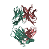 6fabS S: Starting model for refinement |
|---|---|
| Similar structure data |


-Links
 PDBj
PDBj

- Assembly
| Deposited unit | 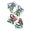
| ||||||||
|---|---|---|---|---|---|---|---|---|---|
| 1 | 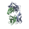
| ||||||||
| 2 | 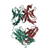
| ||||||||
| Unit cell |
|
-Components
| #1: Antibody | Mass: 23632.947 Da / Num. of mol.: 2 / Fragment: FAB FRAGMENT LIGHT CHAIN / Source method: isolated from a natural source Details: MONOCLONAL ANTIBODY AGAINST ALPHA-D-MANNOPYRANOSIDE Source: (natural) #2: Antibody | Mass: 23789.480 Da / Num. of mol.: 2 / Fragment: FAB FRAGMENT HEAVY CHAIN / Source method: isolated from a natural source Details: MONOCLONAL ANTIBODY AGAINST ALPHA-D-MANNOPYRANOSIDE Source: (natural) #3: Water | ChemComp-HOH / |  Mass: 18.015 Da / Num. of mol.: 106 / Source method: isolated from a natural source / Formula: H2O Mass: 18.015 Da / Num. of mol.: 106 / Source method: isolated from a natural source / Formula: H2OHas protein modification | Y | |
|---|
-Experimental details
-Experiment
| Experiment | Method: X-RAY DIFFRACTION / Number of used crystals: 1 |
|---|
- Sample preparation
| Crystal | Density Matthews: 2.2 Å3/Da / Density % sol: 43.5 % / Description: NONE |
|---|---|
| Crystal grow | pH: 7.4 / Details: 50MM TRISCL, PH 7.4 WITH 20% PEG 4000 |
-Data collection
| Diffraction | Mean temperature: 120 K |
|---|---|
| Diffraction source | Source: ROTATING ANODE / Type: RIGAKU RU300 / Wavelength: 1.5418 |
| Detector | Type: MARRESEARCH / Detector: IMAGE PLATE / Date: Nov 20, 2003 / Details: MIRRORS |
| Radiation | Protocol: SINGLE WAVELENGTH / Monochromatic (M) / Laue (L): M / Scattering type: x-ray |
| Radiation wavelength | Wavelength: 1.5418 Å / Relative weight: 1 |
| Reflection | Resolution: 2.8→50 Å / Num. obs: 19865 / % possible obs: 95.9 % / Observed criterion σ(I): 0 / Redundancy: 3.25 % / Biso Wilson estimate: 38.9 Å2 / Rmerge(I) obs: 0.1 / Net I/σ(I): 7.3 |
| Reflection shell | Resolution: 2.8→2.9 Å / Redundancy: 3.23 % / Rmerge(I) obs: 0.38 / Mean I/σ(I) obs: 1.2 / % possible all: 94.4 |
- Processing
| Software |
| ||||||||||||||||||||||||||||||||||||||||||||||||||||||||||||||||||||||||||||||||
|---|---|---|---|---|---|---|---|---|---|---|---|---|---|---|---|---|---|---|---|---|---|---|---|---|---|---|---|---|---|---|---|---|---|---|---|---|---|---|---|---|---|---|---|---|---|---|---|---|---|---|---|---|---|---|---|---|---|---|---|---|---|---|---|---|---|---|---|---|---|---|---|---|---|---|---|---|---|---|---|---|---|
| Refinement | Method to determine structure: MOLECULAR REPLACEMENT Starting model: PDB ENTRY 6FAB Resolution: 2.8→17.46 Å / Rfactor Rfree error: 0.006 / Data cutoff high absF: 10000 / Isotropic thermal model: RESTRAINED / Cross valid method: THROUGHOUT / σ(F): 0 / Stereochemistry target values: MLF Details: DISORDERED REGIONS IN THE PROTEIN HAVE NOT BEEN MODELED
| ||||||||||||||||||||||||||||||||||||||||||||||||||||||||||||||||||||||||||||||||
| Solvent computation | Solvent model: FLAT MODEL / Bsol: 34.0601 Å2 / ksol: 0.290378 e/Å3 | ||||||||||||||||||||||||||||||||||||||||||||||||||||||||||||||||||||||||||||||||
| Displacement parameters | Biso mean: 45.68 Å2
| ||||||||||||||||||||||||||||||||||||||||||||||||||||||||||||||||||||||||||||||||
| Refine analyze |
| ||||||||||||||||||||||||||||||||||||||||||||||||||||||||||||||||||||||||||||||||
| Refinement step | Cycle: LAST / Resolution: 2.8→17.46 Å
| ||||||||||||||||||||||||||||||||||||||||||||||||||||||||||||||||||||||||||||||||
| Refine LS restraints |
| ||||||||||||||||||||||||||||||||||||||||||||||||||||||||||||||||||||||||||||||||
| LS refinement shell | Resolution: 2.8→2.9 Å / Rfactor Rfree error: 0.027 / Total num. of bins used: 10
| ||||||||||||||||||||||||||||||||||||||||||||||||||||||||||||||||||||||||||||||||
| Xplor file |
|
