 Movie
Movie Controller

[English] 日本語
 Yorodumi
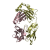
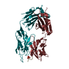
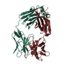
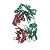
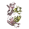
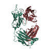
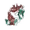
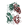
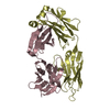
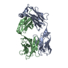
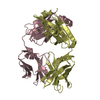
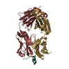
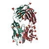
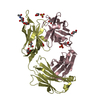
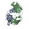
- PDB-1s3k: Crystal Structure of a Humanized Fab (hu3S193) in Complex with th... -
+ Open data

- Basic information
| Entry | Database: PDB / ID: 1s3k | |||||||||
|---|---|---|---|---|---|---|---|---|---|---|
| Title | Crystal Structure of a Humanized Fab (hu3S193) in Complex with the Lewis Y Tetrasaccharide | |||||||||
 Components Components |
| |||||||||
 Keywords Keywords | IMMUNE SYSTEM / Immunoglobulin fold / Beta Barrel / Humanized antibody / Antigen binding fragment | |||||||||
| Function / homology | Immunoglobulins / Immunoglobulin-like / Sandwich / Mainly Beta / Lewis Y antigen, alpha anomer Function and homology information Function and homology information | |||||||||
| Biological species |  | |||||||||
| Method |  X-RAY DIFFRACTION / SYNCHROTRON / MOLECULAR REPLACEMENT / Resolution: 1.9 Å X-RAY DIFFRACTION / SYNCHROTRON / MOLECULAR REPLACEMENT / Resolution: 1.9 Å | |||||||||
 Authors Authors | Ramsland, P.A. / Farrugia, W. / Bradford, T.M. / Hogarth, P.M. / Scott, A.M. | |||||||||
 Citation Citation | Journal: J.Mol.Biol. / Year: 2004 Title: Structural convergence of antibody binding of carbohydrate determinants in lewis y tumor antigens Authors: Ramsland, P.A. / Farrugia, W. / Bradford, T.M. / Hogarth, P.M. / Scott, A.M. | |||||||||
| History |
|
- Structure visualization
| Structure viewer | Molecule: MolmilJmol/JSmol |
|---|
- Downloads & links
-Download
| PDBx/mmCIF format | 1s3k.cif.gz | 106.3 KB | Display | PDBx/mmCIF format |
|---|---|---|---|---|
| PDB format | pdb1s3k.ent.gz | 79 KB | Display | PDB format |
| PDBx/mmJSON format | 1s3k.json.gz | Tree view | PDBx/mmJSON format | |
| Others |  Other downloads Other downloads |
-Validation report
| Arichive directory | https://data.pdbj.org/pub/pdb/validation_reports/s3/1s3kftp://data.pdbj.org/pub/pdb/validation_reports/s3/1s3k | HTTPS FTP |
|---|
-Related structure data
| Related structure data |  1clyS S: Starting model for refinement |
|---|---|
| Similar structure data |


-Links
 PDBj
PDBj

- Assembly
| Deposited unit | 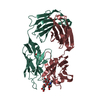
| ||||||||
|---|---|---|---|---|---|---|---|---|---|
| 1 |
| ||||||||
| Unit cell |
|
-Components
| #1: Antibody | Mass: 24066.689 Da / Num. of mol.: 1 Source method: isolated from a genetically manipulated source Details: murine complementarity determining regions grafted onto human framework and constant sequences Source: (gene. exp.) |
|---|---|
| #2: Antibody | Mass: 23796.654 Da / Num. of mol.: 1 Source method: isolated from a genetically manipulated source Details: murine complementarity determining regions grafted onto human framework and constant sequences Source: (gene. exp.) |
| #3: Polysaccharide | alpha-L-fucopyranose-(1-2)-beta-D-galactopyranose-(1-4)-[alpha-L-fucopyranose-(1-3)]2-acetamido-2- ...alpha-L-fucopyranose-(1-2)-beta-D-galactopyranose-(1-4)-[alpha-L-fucopyranose-(1-3)]2-acetamido-2-deoxy-alpha-D-glucopyranose / Lewis Y antigen / alpha anomer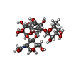  Source method: isolated from a genetically manipulated source Details: oligosaccharide with branches / References: Lewis Y antigen, alpha anomer |
| #4: Chemical | ChemComp-GOL /   Mass: 92.094 Da / Num. of mol.: 1 / Source method: obtained synthetically / Formula: C3H8O3 Mass: 92.094 Da / Num. of mol.: 1 / Source method: obtained synthetically / Formula: C3H8O3 |
| #5: Water | ChemComp-HOH /  Mass: 18.015 Da / Num. of mol.: 346 / Source method: isolated from a natural source / Formula: H2O Mass: 18.015 Da / Num. of mol.: 346 / Source method: isolated from a natural source / Formula: H2O |
| Has protein modification | Y |
-Experimental details
-Experiment
| Experiment | Method: X-RAY DIFFRACTION / Number of used crystals: 1 |
|---|
- Sample preparation
| Crystal | Density Matthews: 2.6 Å3/Da / Density % sol: 52.35 % |
|---|---|
| Crystal grow | Temperature: 291 K / Method: vapor diffusion, sitting drop / pH: 6.5 Details: PEG 20000, sodium chloride, Tris, MES, pH 6.5, VAPOR DIFFUSION, SITTING DROP, temperature 291K |
-Data collection
| Diffraction | Mean temperature: 100 K |
|---|---|
| Diffraction source | Source: SYNCHROTRON / Site: APS  / Beamline: 14-BM-C / Wavelength: 0.9 Å / Beamline: 14-BM-C / Wavelength: 0.9 Å |
| Detector | Type: ADSC QUANTUM 4 / Detector: CCD / Date: Aug 2, 2003 |
| Radiation | Protocol: SINGLE WAVELENGTH / Monochromatic (M) / Laue (L): M / Scattering type: x-ray |
| Radiation wavelength | Wavelength: 0.9 Å / Relative weight: 1 |
| Reflection | Resolution: 1.9→50 Å / Num. all: 39562 / Num. obs: 39562 / % possible obs: 96.5 % / Observed criterion σ(F): 0 / Observed criterion σ(I): 0 / Redundancy: 4.6 % / Biso Wilson estimate: 12.7 Å2 / Rsym value: 0.062 / Net I/σ(I): 15.2 |
| Reflection shell | Resolution: 1.9→1.97 Å / Redundancy: 1.7 % / Mean I/σ(I) obs: 4.8 / Num. unique all: 3008 / Rsym value: 0.153 / % possible all: 73.5 |
- Processing
| Software |
| |||||||||||||||||||||||||
|---|---|---|---|---|---|---|---|---|---|---|---|---|---|---|---|---|---|---|---|---|---|---|---|---|---|---|
| Refinement | Method to determine structure: MOLECULAR REPLACEMENT Starting model: PDB ENTRY 1CLY Resolution: 1.9→33.34 Å / Cross valid method: THROUGHOUT / σ(F): 0 / Stereochemistry target values: Engh & Huber
| |||||||||||||||||||||||||
| Displacement parameters | Biso mean: 27.6 Å2 | |||||||||||||||||||||||||
| Refine analyze |
| |||||||||||||||||||||||||
| Refinement step | Cycle: LAST / Resolution: 1.9→33.34 Å
| |||||||||||||||||||||||||
| Refine LS restraints |
|
