 Movie
Movie Controller

[English] 日本語
 Yorodumi
- PDB-2xkn: Crystal structure of the Fab fragment of the anti-EGFR antibody 7A7 -
+ Open data

- Basic information
| Entry | Database: PDB / ID: 2xkn | ||||||
|---|---|---|---|---|---|---|---|
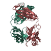
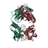
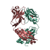
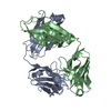
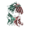
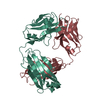
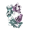
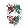
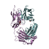
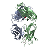
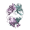
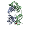
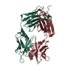
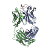
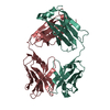
| Title | Crystal structure of the Fab fragment of the anti-EGFR antibody 7A7 | ||||||
 Components Components | (ANTI-EGFR ANTIBODY 7A7) x 2 | ||||||
 Keywords Keywords | IMMUNE SYSTEM / IMMUNOGLOBULIN | ||||||
| Function / homology | Immunoglobulins / Immunoglobulin-like / Sandwich / Mainly Beta Function and homology information Function and homology information | ||||||
| Biological species |  | ||||||
| Method |  X-RAY DIFFRACTION / SYNCHROTRON / MOLECULAR REPLACEMENT / Resolution: 1.4 Å X-RAY DIFFRACTION / SYNCHROTRON / MOLECULAR REPLACEMENT / Resolution: 1.4 Å | ||||||
 Authors Authors | Talavera, A. / Mackenzie, J. / Friemann, R. / Krengel, U. | ||||||
 Citation Citation | Journal: Mol.Immunol. / Year: 2011 Title: Structure of the Fab Fragment of the Anti-Murine Egfr Antibody 7A7 and Exploration of its Receptor Binding Site. Authors: Talavera, A. / Mackenzie, J. / Garrido, G. / Friemann, R. / Lopez-Requena, A. / Moreno, E. / Krengel, U. | ||||||
| History |
|
- Structure visualization
| Structure viewer | Molecule: MolmilJmol/JSmol |
|---|
- Downloads & links
-Download
| PDBx/mmCIF format | 2xkn.cif.gz | 396.1 KB | Display | PDBx/mmCIF format |
|---|---|---|---|---|
| PDB format | pdb2xkn.ent.gz | 326.6 KB | Display | PDB format |
| PDBx/mmJSON format | 2xkn.json.gz | Tree view | PDBx/mmJSON format | |
| Others |  Other downloads Other downloads |
-Validation report
| Arichive directory | https://data.pdbj.org/pub/pdb/validation_reports/xk/2xknftp://data.pdbj.org/pub/pdb/validation_reports/xk/2xkn | HTTPS FTP |
|---|
-Related structure data
| Related structure data |  1hilS S: Starting model for refinement |
|---|---|
| Similar structure data |


-Links
 PDBj
PDBj

- Assembly
| Deposited unit | 
| |||||||||||||||
|---|---|---|---|---|---|---|---|---|---|---|---|---|---|---|---|---|
| 1 | 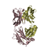
| |||||||||||||||
| 2 | 
| |||||||||||||||
| Unit cell |
| |||||||||||||||
| Components on special symmetry positions |
|
-Components
| #1: Antibody | Mass: 24628.053 Da / Num. of mol.: 2 / Fragment: FAB FRAGMENT LIGHT CHAIN (IGG1), RESIDUES 1-223 / Source method: isolated from a natural source / Details: HYBRIDOMA / Source: (natural) #2: Antibody | Mass: 23124.041 Da / Num. of mol.: 2 / Fragment: FAB FRAGMENT HEAVY CHAIN (IGG1), RESIDUES 1-216 / Source method: isolated from a natural source / Details: HYBRIDOMA / Source: (natural) #3: Chemical | ChemComp-PE4 /   Mass: 354.436 Da / Num. of mol.: 4 / Source method: obtained synthetically / Formula: C16H34O8 / Comment: precipitant*YM Mass: 354.436 Da / Num. of mol.: 4 / Source method: obtained synthetically / Formula: C16H34O8 / Comment: precipitant*YM#4: Water | ChemComp-HOH / |  Mass: 18.015 Da / Num. of mol.: 1112 / Source method: isolated from a natural source / Formula: H2O Mass: 18.015 Da / Num. of mol.: 1112 / Source method: isolated from a natural source / Formula: H2OHas protein modification | Y | |
|---|
-Experimental details
-Experiment
| Experiment | Method: X-RAY DIFFRACTION / Number of used crystals: 1 |
|---|
- Sample preparation
| Crystal | Density Matthews: 2.5 Å3/Da / Density % sol: 50 % / Description: NONE |
|---|---|
| Crystal grow | Details: 15% PEG 8000, 100 MM TRISAC BUFFER PH 9.0, 10 MM EDTA FROM THE HAMPTON ADDITIVE SCREEN |
-Data collection
| Diffraction | Mean temperature: 90 K |
|---|---|
| Diffraction source | Source: SYNCHROTRON / Site: ESRF  / Beamline: ID14-3 / Wavelength: 0.931 / Beamline: ID14-3 / Wavelength: 0.931 |
| Detector | Detector: CCD / Date: May 16, 2007 / Details: MIRRORS |
| Radiation | Monochromator: DIAMOND (111) / Protocol: SINGLE WAVELENGTH / Monochromatic (M) / Laue (L): M / Scattering type: x-ray |
| Radiation wavelength | Wavelength: 0.931 Å / Relative weight: 1 |
| Reflection | Resolution: 1.4→23 Å / Num. obs: 191075 / % possible obs: 99.6 % / Observed criterion σ(I): 2 / Redundancy: 4.5 % / Biso Wilson estimate: 14.77 Å2 / Rmerge(I) obs: 0.06 / Net I/σ(I): 13.1 |
| Reflection shell | Resolution: 1.4→1.42 Å / Redundancy: 4.1 % / Rmerge(I) obs: 0.36 / Mean I/σ(I) obs: 3.1 / % possible all: 98.9 |
- Processing
| Software |
| |||||||||||||||||||||||||||||||||||||||||||||||||||||||||||||||||||||||||||||||||||||||||||||||||||||||||||||||||||||||||||||||||||||||||||||||||||||||||||||||||||||||||||||||||||||||||||||||||||||||||||||||||||||||||
|---|---|---|---|---|---|---|---|---|---|---|---|---|---|---|---|---|---|---|---|---|---|---|---|---|---|---|---|---|---|---|---|---|---|---|---|---|---|---|---|---|---|---|---|---|---|---|---|---|---|---|---|---|---|---|---|---|---|---|---|---|---|---|---|---|---|---|---|---|---|---|---|---|---|---|---|---|---|---|---|---|---|---|---|---|---|---|---|---|---|---|---|---|---|---|---|---|---|---|---|---|---|---|---|---|---|---|---|---|---|---|---|---|---|---|---|---|---|---|---|---|---|---|---|---|---|---|---|---|---|---|---|---|---|---|---|---|---|---|---|---|---|---|---|---|---|---|---|---|---|---|---|---|---|---|---|---|---|---|---|---|---|---|---|---|---|---|---|---|---|---|---|---|---|---|---|---|---|---|---|---|---|---|---|---|---|---|---|---|---|---|---|---|---|---|---|---|---|---|---|---|---|---|---|---|---|---|---|---|---|---|---|---|---|---|---|---|---|---|
| Refinement | Method to determine structure: MOLECULAR REPLACEMENT Starting model: PDB ENTRY 1HIL Resolution: 1.4→22.614 Å / SU ML: 0.19 / σ(F): 1.12 / Phase error: 15.8 / Stereochemistry target values: ML
| |||||||||||||||||||||||||||||||||||||||||||||||||||||||||||||||||||||||||||||||||||||||||||||||||||||||||||||||||||||||||||||||||||||||||||||||||||||||||||||||||||||||||||||||||||||||||||||||||||||||||||||||||||||||||
| Solvent computation | Shrinkage radii: 0.9 Å / VDW probe radii: 1.11 Å / Solvent model: FLAT BULK SOLVENT MODEL / Bsol: 46.638 Å2 / ksol: 0.376 e/Å3 | |||||||||||||||||||||||||||||||||||||||||||||||||||||||||||||||||||||||||||||||||||||||||||||||||||||||||||||||||||||||||||||||||||||||||||||||||||||||||||||||||||||||||||||||||||||||||||||||||||||||||||||||||||||||||
| Displacement parameters | Biso mean: 20 Å2
| |||||||||||||||||||||||||||||||||||||||||||||||||||||||||||||||||||||||||||||||||||||||||||||||||||||||||||||||||||||||||||||||||||||||||||||||||||||||||||||||||||||||||||||||||||||||||||||||||||||||||||||||||||||||||
| Refinement step | Cycle: LAST / Resolution: 1.4→22.614 Å
| |||||||||||||||||||||||||||||||||||||||||||||||||||||||||||||||||||||||||||||||||||||||||||||||||||||||||||||||||||||||||||||||||||||||||||||||||||||||||||||||||||||||||||||||||||||||||||||||||||||||||||||||||||||||||
| Refine LS restraints |
| |||||||||||||||||||||||||||||||||||||||||||||||||||||||||||||||||||||||||||||||||||||||||||||||||||||||||||||||||||||||||||||||||||||||||||||||||||||||||||||||||||||||||||||||||||||||||||||||||||||||||||||||||||||||||
| LS refinement shell |
|
