#62 - Feb 2005 Major Histocompatibility Complex similarity (7)
#170 - Feb 2014 Broadly Neutralizing Antibodies similarity (6)
#252 - Dec 2020 Hepatitis C Virus Protease/Helicase similarity (1)
-
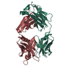
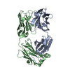
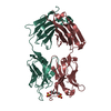
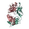
Assembly
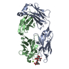
Deposited unit
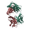
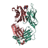
L: Light chain of Fab fragment of rabbit anti-HIV1 gp120 V3 mAb 10A3 H: Heavy chain of Fab fragment of rabbit anti-HIV1 gp120 V3 mAb 10A3 P: Envelope glycoprotein gp120 V3 peptide of Con B sequence hetero molecules
Mass: 23384.846 Da / Num. of mol.: 1 Source method: isolated from a genetically manipulated source Source: (gene. exp.) Oryctolagus cuniculus (rabbit) / Production host: Homo sapiens (human)
Mass: 22305.047 Da / Num. of mol.: 1 Source method: isolated from a genetically manipulated source Source: (gene. exp.) Oryctolagus cuniculus (rabbit) / Production host: Homo sapiens
#3: Protein/peptide
Envelopeglycoproteingp120V3peptideofConBsequence
Mass: 1671.902 Da / Num. of mol.: 1 / Source method: obtained synthetically / Source: (synth.) Human immunodeficiency virus 1 / References: UniProt: P20871*PLUS
In the structure databanks used in Yorodumi, some data are registered as the other names, "COVID-19 virus" and "2019-nCoV". Here are the details of the virus and the list of structure data.
Jan 31, 2019. EMDB accession codes are about to change! (news from PDBe EMDB page)
EMDB accession codes are about to change! (news from PDBe EMDB page)
The allocation of 4 digits for EMDB accession codes will soon come to an end. Whilst these codes will remain in use, new EMDB accession codes will include an additional digit and will expand incrementally as the available range of codes is exhausted. The current 4-digit format prefixed with “EMD-” (i.e. EMD-XXXX) will advance to a 5-digit format (i.e. EMD-XXXXX), and so on. It is currently estimated that the 4-digit codes will be depleted around Spring 2019, at which point the 5-digit format will come into force.
The EM Navigator/Yorodumi systems omit the EMD- prefix.
Related info.:Q: What is EMD? / ID/Accession-code notation in Yorodumi/EM Navigator
Yorodumi is a browser for structure data from EMDB, PDB, SASBDB, etc.
This page is also the successor to EM Navigator detail page, and also detail information page/front-end page for Omokage search.
The word "yorodu" (or yorozu) is an old Japanese word meaning "ten thousand". "mi" (miru) is to see.
Related info.:EMDB / PDB / SASBDB / Comparison of 3 databanks / Yorodumi Search / Aug 31, 2016. New EM Navigator & Yorodumi / Yorodumi Papers / Jmol/JSmol / Function and homology information / Changes in new EM Navigator and Yorodumi
 Movie
Movie Controller

 Yorodumi
 Open data
 Basic information
 Components
 Keywords
 Function and homology information

 Human immunodeficiency virus 1
 X-RAY DIFFRACTION /
  Authors
 United States, 2items
  Citation
 Structure visualization
 Downloads & links
 Other downloads
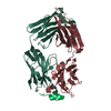


 PDBj
PDBj


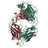
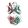
 Assembly
 Homo sapiens (human)
 Mass: 40.078 Da / Num. of mol.: 1 / Source method: obtained synthetically / Formula: Ca
 Mass: 18.015 Da / Num. of mol.: 668 / Source method: isolated from a natural source / Formula: H2O
 Sample preparation
 Processing