 Movie
Movie Controller

[English] 日本語
 Yorodumi
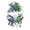
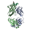
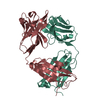
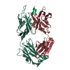
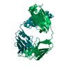
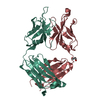
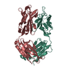
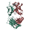
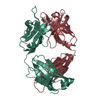
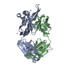
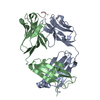
- PDB-4hha: Anti-Human Cytomegalovirus (HCMV) Fab KE5 with epitope peptide AD-2S1 -
+ Open data

- Basic information
| Entry | Database: PDB / ID: 4hha | ||||||
|---|---|---|---|---|---|---|---|
| Title | Anti-Human Cytomegalovirus (HCMV) Fab KE5 with epitope peptide AD-2S1 | ||||||
 Components Components |
| ||||||
 Keywords Keywords | IMMUNE SYSTEM / Fab fragment / Antibody | ||||||
| Function / homology | Herpesvirus Glycoprotein B, antigenic domain, N-terminal / Glycoprotein B N-terminal antigenic domain of HCMV / Immunoglobulins / Immunoglobulin-like / Sandwich / Mainly Beta / Glycoprotein B Function and homology information Function and homology information | ||||||
| Biological species |  Homo sapiens (human) Homo sapiens (human)  Human herpesvirus 5 Human herpesvirus 5 | ||||||
| Method |  X-RAY DIFFRACTION / SYNCHROTRON / MOLECULAR REPLACEMENT / Resolution: 1.6 Å X-RAY DIFFRACTION / SYNCHROTRON / MOLECULAR REPLACEMENT / Resolution: 1.6 Å | ||||||
 Authors Authors | Bryson, S. / Risnes, L. / Damgupta, S. / Thomson, C.A. / Pfoh, R. / Schrader, J.W. / Pai, E.F. | ||||||
 Citation Citation | Journal: J. Immunol. / Year: 2016 Title: Structures of Preferred Human IgV Genes-Based Protective Antibodies Identify How Conserved Residues Contact Diverse Antigens and Assign Source of Specificity to CDR3 Loop Variation. Authors: Bryson, S. / Thomson, C.A. / Risnes, L.F. / Dasgupta, S. / Smith, K. / Schrader, J.W. / Pai, E.F. | ||||||
| History |
|
- Structure visualization
| Structure viewer | Molecule: MolmilJmol/JSmol |
|---|
- Downloads & links
-Download
| PDBx/mmCIF format | 4hha.cif.gz | 104.6 KB | Display | PDBx/mmCIF format |
|---|---|---|---|---|
| PDB format | pdb4hha.ent.gz | 78.4 KB | Display | PDB format |
| PDBx/mmJSON format | 4hha.json.gz | Tree view | PDBx/mmJSON format | |
| Others |  Other downloads Other downloads |
-Validation report
| Arichive directory | https://data.pdbj.org/pub/pdb/validation_reports/hh/4hhaftp://data.pdbj.org/pub/pdb/validation_reports/hh/4hha | HTTPS FTP |
|---|
-Related structure data
| Related structure data |  4hh9SC  4hieC 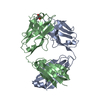 4hihC 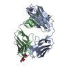 4hiiC 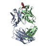 4hijC  4pttC 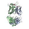 4ptuC S: Starting model for refinement C: citing same article ( |
|---|---|
| Similar structure data |


-Links
 PDBj
PDBj

- Assembly
| Deposited unit | 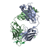
| ||||||||
|---|---|---|---|---|---|---|---|---|---|
| 1 |
| ||||||||
| Unit cell |
|
-Components
| #1: Antibody | Mass: 23471.064 Da / Num. of mol.: 1 / Fragment: Fab fragment KE5 / Source method: isolated from a natural source / Source: (natural) Homo sapiens (human) | ||||
|---|---|---|---|---|---|
| #2: Antibody | Mass: 25400.516 Da / Num. of mol.: 1 / Fragment: AD-2S1 eptiope / Source method: isolated from a natural source / Source: (natural) Homo sapiens (human) | ||||
| #3: Protein/peptide | Mass: 1246.386 Da / Num. of mol.: 1 / Source method: obtained synthetically / Details: synthesized peptide / Source: (synth.) Human herpesvirus 5 / References: UniProt: Q9YKI2 | ||||
| #4: Chemical | ChemComp-CL /   Mass: 35.453 Da / Num. of mol.: 5 / Source method: obtained synthetically / Formula: Cl Mass: 35.453 Da / Num. of mol.: 5 / Source method: obtained synthetically / Formula: Cl#5: Water | ChemComp-HOH / |  Mass: 18.015 Da / Num. of mol.: 289 / Source method: isolated from a natural source / Formula: H2O Mass: 18.015 Da / Num. of mol.: 289 / Source method: isolated from a natural source / Formula: H2OHas protein modification | Y | |
-Experimental details
-Experiment
| Experiment | Method: X-RAY DIFFRACTION / Number of used crystals: 1 |
|---|
- Sample preparation
| Crystal | Density Matthews: 1.88 Å3/Da / Density % sol: 34.73 % |
|---|---|
| Crystal grow | Temperature: 298 K / Method: vapor diffusion, hanging drop / pH: 4.2 Details: 0.05M Na Acetate, pH 4.2, 18% Jeffamine 1500, 0.2M Ammonium chloride , VAPOR DIFFUSION, HANGING DROP, temperature 298.0K |
-Data collection
| Diffraction | Mean temperature: 110 K |
|---|---|
| Diffraction source | Source: SYNCHROTRON / Site: CLSI  / Beamline: 08ID-1 / Wavelength: 1.0332 Å / Beamline: 08ID-1 / Wavelength: 1.0332 Å |
| Detector | Type: RAYONIX MX300HE / Detector: CCD / Date: Sep 28, 2012 / Details: Toroidal focusing mirror |
| Radiation | Monochromator: DCM / Protocol: SINGLE WAVELENGTH / Monochromatic (M) / Laue (L): M / Scattering type: x-ray |
| Radiation wavelength | Wavelength: 1.0332 Å / Relative weight: 1 |
| Reflection | Resolution: 1.6→43 Å / Num. all: 49164 / Num. obs: 48944 / % possible obs: 99.6 % / Observed criterion σ(F): 0 / Observed criterion σ(I): 0 / Redundancy: 3.72 % / Rmerge(I) obs: 0.0478 / Net I/σ(I): 19.84 |
| Reflection shell | Resolution: 1.6→1.65 Å / Redundancy: 3.69 % / Rmerge(I) obs: 0.492 / Mean I/σ(I) obs: 2.89 / Num. unique all: 4260 / % possible all: 99.9 |
- Processing
| Software |
| ||||||||||||||||||||
|---|---|---|---|---|---|---|---|---|---|---|---|---|---|---|---|---|---|---|---|---|---|
| Refinement | Method to determine structure: MOLECULAR REPLACEMENT Starting model: PDB ENTRY 4HH9 Resolution: 1.6→43 Å / σ(F): 0 / Stereochemistry target values: Engh & Huber
| ||||||||||||||||||||
| Refinement step | Cycle: LAST / Resolution: 1.6→43 Å
| ||||||||||||||||||||
| Refine LS restraints |
|
