 Movie
Movie Controller


+ Open data

- Basic information
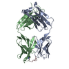
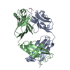
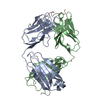
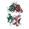
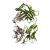
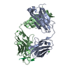
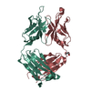
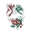
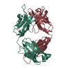
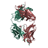
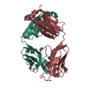
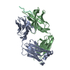
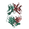
| Entry | Database: PDB / ID: 1hi6 | ||||||
|---|---|---|---|---|---|---|---|
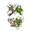
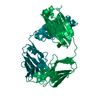
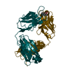
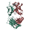
| Title | ANTI-P24 (HIV-1) FAB FRAGMENT CB41 COMPLEXED WITH A PEPTIDE | ||||||
 Components Components |
| ||||||
 Keywords Keywords | IMMUNE SYSTEM/PEPTIDE / COMPLEX (ANTIBODY-PEPTIDE) / POLYSPECIFICITY / CROSSREACTIVITY / FAB-FRAGMENT / HIV-1 / IMMUNE SYSTEM-PEPTIDE complex | ||||||
| Function / homology |  Function and homology information Function and homology informationalpha-beta T cell receptor complex / immunoglobulin complex, circulating / immunoglobulin receptor binding / IgG immunoglobulin complex / complement activation, classical pathway / antigen binding / B cell differentiation / antibacterial humoral response / adaptive immune response / extracellular region / plasma membrane Similarity search - Function | ||||||
| Biological species |  SYNTHETIC CONSTRUCT (others) | ||||||
| Method |  X-RAY DIFFRACTION / SYNCHROTRON / MOLECULAR REPLACEMENT / Resolution: 2.55 Å X-RAY DIFFRACTION / SYNCHROTRON / MOLECULAR REPLACEMENT / Resolution: 2.55 Å | ||||||
 Authors Authors | Hahn, M. / Wessner, H. / Schneider-Mergener, J. / Hohne, W. | ||||||
 Citation Citation | Journal: Cell(Cambridge,Mass.) / Year: 1997 Title: Crystallographic Analysis of Anti-P24 (HIV-1) Monoclonal Antibody Cross-Reactivity and Polyspecificity Authors: Keitel, T. / Kramer, A. / Wessner, H. / Scholz, C. / Schneider-Mergener, J. / Hohne, W. | ||||||
| History |
|
- Structure visualization
| Structure viewer | Molecule: MolmilJmol/JSmol |
|---|
- Downloads & links
-Download
| PDBx/mmCIF format | 1hi6.cif.gz | 97.5 KB | Display | PDBx/mmCIF format |
|---|---|---|---|---|
| PDB format | pdb1hi6.ent.gz | 75 KB | Display | PDB format |
| PDBx/mmJSON format | 1hi6.json.gz | Tree view | PDBx/mmJSON format | |
| Others |  Other downloads Other downloads |
-Validation report
| Arichive directory | https://data.pdbj.org/pub/pdb/validation_reports/hi/1hi6ftp://data.pdbj.org/pub/pdb/validation_reports/hi/1hi6 | HTTPS FTP |
|---|
-Related structure data
| Related structure data |  1bogSC 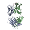 1cfnC 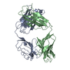 1cfqC 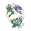 1cfsC 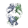 1cftC S: Starting model for refinement C: citing same article ( |
|---|---|
| Similar structure data |


-Links
 PDBj
PDBj


- Assembly
| Deposited unit | 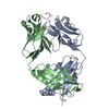
| ||||||||
|---|---|---|---|---|---|---|---|---|---|
| 1 |
| ||||||||
| Unit cell |
|
-Components
| #1: Antibody | Mass: 23928.721 Da / Num. of mol.: 1 / Source method: isolated from a natural source / Source: (natural) |
|---|---|
| #2: Antibody | Mass: 22669.508 Da / Num. of mol.: 1 / Source method: isolated from a natural source / Source: (natural) |
| #3: Protein/peptide | Mass: 1227.370 Da / Num. of mol.: 1 / Source method: obtained synthetically / Details: EPITOPE-RELATED PEPTIDE / Source: (synth.) SYNTHETIC CONSTRUCT (others) |
| #4: Water | ChemComp-HOH /  Mass: 18.015 Da / Num. of mol.: 59 / Source method: isolated from a natural source / Formula: H2O Mass: 18.015 Da / Num. of mol.: 59 / Source method: isolated from a natural source / Formula: H2O |
| Compound details | NH2: THE SYNTHETIC PEPTIDE HAS ITS C-TERMINAL END BLOCKED WITH AN AMIDE GROUP. |
| Has protein modification | Y |
| Sequence details | THE SEQUENCE IS RELATED TO THE ENTRY 1BOG. THE TWO RESIDUES IN THIS ENTRY VALB168, THRB171 ARE ...THE SEQUENCE IS RELATED TO THE ENTRY 1BOG. THE TWO RESIDUES IN THIS ENTRY VALB168, THRB171 ARE LEU168 AND SER171 RESPECTIVE |
-Experimental details
-Experiment
| Experiment | Method: X-RAY DIFFRACTION / Number of used crystals: 1 |
|---|
- Sample preparation
| Crystal | Density Matthews: 4.7 Å3/Da / Density % sol: 71 % |
|---|---|
| Crystal grow | Method: vapor diffusion, hanging drop / pH: 7.5 Details: HANGING DROP SET UP AT ROOM TEMPERATURE. PROTEIN CONCENTRATION: 10 MG/ML RESERVOIR: 1.8 M AMMONIUM SULFATE, 0.1 M MOPS-BUFFER, PH 7.5. |
| Crystal grow | *PLUS Method: unknown |
-Data collection
| Diffraction | Mean temperature: 100 K |
|---|---|
| Diffraction source | Source: SYNCHROTRON / Site: EMBL/DESY, HAMBURG  / Beamline: X11 / Wavelength: 0.9 / Beamline: X11 / Wavelength: 0.9 |
| Detector | Type: MARRESEARCH / Detector: IMAGE PLATE / Date: Apr 15, 1999 |
| Radiation | Protocol: SINGLE WAVELENGTH / Monochromatic (M) / Laue (L): M / Scattering type: x-ray |
| Radiation wavelength | Wavelength: 0.9 Å / Relative weight: 1 |
| Reflection | Resolution: 2.55→90 Å / Num. obs: 2976 / % possible obs: 95.4 % / Redundancy: 4.8 % / Rmerge(I) obs: 0.088 / Net I/σ(I): 24.8 |
| Reflection shell | Resolution: 2.55→2.64 Å / Redundancy: 5.2 % / Rmerge(I) obs: 0.401 / Mean I/σ(I) obs: 3.8 / % possible all: 98.6 |
- Processing
| Software |
| ||||||||||||||||||||||||||||||||||||||||||||||||||||||||||||
|---|---|---|---|---|---|---|---|---|---|---|---|---|---|---|---|---|---|---|---|---|---|---|---|---|---|---|---|---|---|---|---|---|---|---|---|---|---|---|---|---|---|---|---|---|---|---|---|---|---|---|---|---|---|---|---|---|---|---|---|---|---|
| Refinement | Method to determine structure: MOLECULAR REPLACEMENT Starting model: 1BOG Resolution: 2.55→20 Å / Isotropic thermal model: RESTRAINED / Cross valid method: THROUGHOUT / σ(F): 0
| ||||||||||||||||||||||||||||||||||||||||||||||||||||||||||||
| Displacement parameters | Biso mean: 44.66 Å2
| ||||||||||||||||||||||||||||||||||||||||||||||||||||||||||||
| Refinement step | Cycle: LAST / Resolution: 2.55→20 Å
| ||||||||||||||||||||||||||||||||||||||||||||||||||||||||||||
| Refine LS restraints |
| ||||||||||||||||||||||||||||||||||||||||||||||||||||||||||||
| LS refinement shell | Resolution: 2.55→2.58 Å / Total num. of bins used: 29
| ||||||||||||||||||||||||||||||||||||||||||||||||||||||||||||
| Xplor file |
|
