 Movie
Movie Controller

[English] 日本語
 Yorodumi
- PDB-3pp4: Epitope characterization and crystal structure of GA101 provide i... -
+ Open data

- Basic information
| Entry | Database: PDB / ID: 3pp4 | ||||||
|---|---|---|---|---|---|---|---|
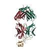
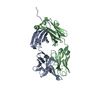
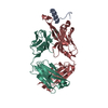
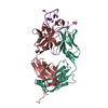
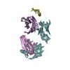
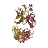
| Title | Epitope characterization and crystal structure of GA101 provide insights into the molecular basis for the type I / type II distinction of anti- CD20 antibodies | ||||||
 Components Components |
| ||||||
 Keywords Keywords | IMMUNE SYSTEM / antibody Fab fragment Ig-domain / CD20 / cyclic peptide / antibody antigen | ||||||
| Function / homology |  Function and homology information Function and homology informationstore-operated calcium entry / calcium ion import into cytosol / positive regulation of calcium ion import across plasma membrane / epidermal growth factor receptor binding / humoral immune response / B cell proliferation / B cell activation / immunoglobulin binding / plasma membrane raft / B cell differentiation ...store-operated calcium entry / calcium ion import into cytosol / positive regulation of calcium ion import across plasma membrane / epidermal growth factor receptor binding / humoral immune response / B cell proliferation / B cell activation / immunoglobulin binding / plasma membrane raft / B cell differentiation / B cell receptor signaling pathway / response to bacterium / protein tetramerization / MHC class II protein complex binding / cell surface receptor signaling pathway / external side of plasma membrane / cell surface / : / extracellular exosome / nucleoplasm / identical protein binding / plasma membrane Similarity search - Function | ||||||
| Biological species |   Homo sapiens (human) Homo sapiens (human) | ||||||
| Method |  X-RAY DIFFRACTION / SYNCHROTRON / MOLECULAR REPLACEMENT / Resolution: 1.6 Å X-RAY DIFFRACTION / SYNCHROTRON / MOLECULAR REPLACEMENT / Resolution: 1.6 Å | ||||||
 Authors Authors | Hopfner, K.-P. / Lammens, A. | ||||||
 Citation Citation | Journal: Blood / Year: 2011 Title: Epitope characterization and crystal structure of GA101 provide insights into the molecular basis for type I/II distinction of CD20 antibodies. Authors: Niederfellner, G. / Lammens, A. / Mundigl, O. / Georges, G.J. / Schaefer, W. / Schwaiger, M. / Franke, A. / Wiechmann, K. / Jenewein, S. / Slootstra, J.W. / Timmerman, P. / Brannstrom, A. / ...Authors: Niederfellner, G. / Lammens, A. / Mundigl, O. / Georges, G.J. / Schaefer, W. / Schwaiger, M. / Franke, A. / Wiechmann, K. / Jenewein, S. / Slootstra, J.W. / Timmerman, P. / Brannstrom, A. / Lindstrom, F. / Mossner, E. / Umana, P. / Hopfner, K.P. / Klein, C. | ||||||
| History |
|
- Structure visualization
| Structure viewer | Molecule: MolmilJmol/JSmol |
|---|
- Downloads & links
-Download
| PDBx/mmCIF format | 3pp4.cif.gz | 234.8 KB | Display | PDBx/mmCIF format |
|---|---|---|---|---|
| PDB format | pdb3pp4.ent.gz | 188.3 KB | Display | PDB format |
| PDBx/mmJSON format | 3pp4.json.gz | Tree view | PDBx/mmJSON format | |
| Others |  Other downloads Other downloads |
-Validation report
| Arichive directory | https://data.pdbj.org/pub/pdb/validation_reports/pp/3pp4ftp://data.pdbj.org/pub/pdb/validation_reports/pp/3pp4 | HTTPS FTP |
|---|
-Related structure data
| Related structure data |  3pp3SC S: Starting model for refinement C: citing same article ( |
|---|---|
| Similar structure data |


-Links
 PDBj
PDBj

- Assembly
| Deposited unit | 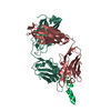
| ||||||||
|---|---|---|---|---|---|---|---|---|---|
| 1 |
| ||||||||
| Unit cell |
|
-Components
| #1: Antibody | Mass: 23969.881 Da / Num. of mol.: 1 Source method: isolated from a genetically manipulated source Source: (gene. exp.)  Cricetulus griseus (Chinese hamster) Cricetulus griseus (Chinese hamster) |
|---|---|
| #2: Antibody | Mass: 23964.822 Da / Num. of mol.: 1 Source method: isolated from a genetically manipulated source Source: (gene. exp.) Cricetulus griseus (Chinese hamster) |
| #3: Protein/peptide | Mass: 2852.051 Da / Num. of mol.: 1 / Fragment: large extracellular loop (UNP residues 163-187) / Source method: obtained synthetically / Details: CD20 / Source: (synth.) Homo sapiens (human) / References: UniProt: P11836 |
| #4: Chemical | ChemComp-CL /   Mass: 35.453 Da / Num. of mol.: 1 / Source method: obtained synthetically / Formula: Cl Mass: 35.453 Da / Num. of mol.: 1 / Source method: obtained synthetically / Formula: Cl |
| #5: Water | ChemComp-HOH /  Mass: 18.015 Da / Num. of mol.: 759 / Source method: isolated from a natural source / Formula: H2O Mass: 18.015 Da / Num. of mol.: 759 / Source method: isolated from a natural source / Formula: H2O |
| Has protein modification | Y |
-Experimental details
-Experiment
| Experiment | Method: X-RAY DIFFRACTION / Number of used crystals: 1 |
|---|
- Sample preparation
| Crystal | Density Matthews: 2.32 Å3/Da / Density % sol: 47 % |
|---|---|
| Crystal grow | Temperature: 293 K / Method: vapor diffusion, hanging drop / pH: 6 Details: 16% (w/v) PEG4000, 4% (v/v) isopropanol, 0.1 M sodium acetate, pH 6.0, VAPOR DIFFUSION, HANGING DROP, temperature 293K |
-Data collection
| Diffraction | Mean temperature: 100 K |
|---|---|
| Diffraction source | Source: SYNCHROTRON / Site: ESRF  / Beamline: ID23-1 / Wavelength: 1 Å / Beamline: ID23-1 / Wavelength: 1 Å |
| Detector | Type: ADSC QUANTUM 315r / Detector: CCD / Date: Nov 13, 2008 |
| Radiation | Monochromator: Silicon (1 1 1) channel-cut / Protocol: SINGLE WAVELENGTH / Monochromatic (M) / Laue (L): M / Scattering type: x-ray |
| Radiation wavelength | Wavelength: 1 Å / Relative weight: 1 |
| Reflection | Resolution: 1.6→50 Å / Num. all: 61332 / Num. obs: 60342 / % possible obs: 98.4 % / Observed criterion σ(F): -3 / Redundancy: 3.7 % / Rmerge(I) obs: 0.055 / Rsym value: 0.043 / Net I/σ(I): 17.9 |
| Reflection shell | Resolution: 1.6→1.7 Å / Redundancy: 3.7 % / Rmerge(I) obs: 0.247 / Mean I/σ(I) obs: 5.9 / Rsym value: 0.21 / % possible all: 96.8 |
- Processing
| Software |
| |||||||||||||||||||||||||||||||||||||||||||||||||||||||||||||||||||||||||||||||||||||||||||||||||||||||||
|---|---|---|---|---|---|---|---|---|---|---|---|---|---|---|---|---|---|---|---|---|---|---|---|---|---|---|---|---|---|---|---|---|---|---|---|---|---|---|---|---|---|---|---|---|---|---|---|---|---|---|---|---|---|---|---|---|---|---|---|---|---|---|---|---|---|---|---|---|---|---|---|---|---|---|---|---|---|---|---|---|---|---|---|---|---|---|---|---|---|---|---|---|---|---|---|---|---|---|---|---|---|---|---|---|---|---|
| Refinement | Method to determine structure: MOLECULAR REPLACEMENT Starting model: 3PP3 Resolution: 1.6→42.95 Å / SU ML: 0.17 / σ(F): 1.97 / Stereochemistry target values: ML
| |||||||||||||||||||||||||||||||||||||||||||||||||||||||||||||||||||||||||||||||||||||||||||||||||||||||||
| Solvent computation | Shrinkage radii: 0.9 Å / VDW probe radii: 1.11 Å / Solvent model: FLAT BULK SOLVENT MODEL / Bsol: 76.508 Å2 / ksol: 0.38 e/Å3 | |||||||||||||||||||||||||||||||||||||||||||||||||||||||||||||||||||||||||||||||||||||||||||||||||||||||||
| Displacement parameters |
| |||||||||||||||||||||||||||||||||||||||||||||||||||||||||||||||||||||||||||||||||||||||||||||||||||||||||
| Refinement step | Cycle: LAST / Resolution: 1.6→42.95 Å
| |||||||||||||||||||||||||||||||||||||||||||||||||||||||||||||||||||||||||||||||||||||||||||||||||||||||||
| Refine LS restraints |
| |||||||||||||||||||||||||||||||||||||||||||||||||||||||||||||||||||||||||||||||||||||||||||||||||||||||||
| LS refinement shell |
|
