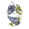
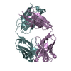
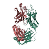
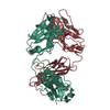
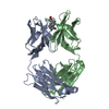
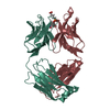
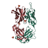
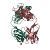
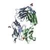
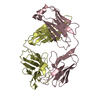
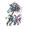
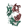
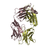
Resolution: 1.78→23 Å / Isotropic thermal model: isotropic / Cross valid method: THROUGHOUT / σ(F): 0 / σ(I): 0 / Stereochemistry target values: Engh & Huber Details: The current model includes the FAB fragment TE33, residues DVA A 1 - DSN A 9 of the bound D-amino acid peptide D2, 276 water molecules and 2 CITRATE ion. The light and heavy chains have been ...Details: The current model includes the FAB fragment TE33, residues DVA A 1 - DSN A 9 of the bound D-amino acid peptide D2, 276 water molecules and 2 CITRATE ion. The light and heavy chains have been designated *L* and *H*, respectively. Peptide has been designated *A*. The numbering of TE33 residues is essentially according to E. KABAT (E.A. KABAT,T.T. WU, H.M. PERRY, K.S. GOTTESMAN, sequences of proteins of Imuunological interest, 5TH ED.,(1991), NATIONAL INSTITUTES OF HEALTH, BETHESDA, MD.). There is no electron density corresponding to the 7-residue stretch H 127 - H 133 in the constant domain of heavy chain H.
Rfactor
Num. reflection
% reflection
Selection details
Rfree
0.248
1989
-
random
Rwork
0.198
-
-
-
all
0.201
40407
-
-
obs
0.201
40407
93.4 %
-
Displacement parameters
Biso mean: 23.0271 Å2
Baniso -1
Baniso -2
Baniso -3
1-
-2.057 Å2
0 Å2
0 Å2
2-
-
0.188 Å2
0 Å2
3-
-
-
1.868 Å2
Refine analyze
Free
Obs
Luzzati coordinate error
0.09 Å
0.21 Å
Luzzati d res low
-
5 Å
Luzzati sigma a
0.13 Å
0.26 Å
Refinement step
Cycle: LAST / Resolution: 1.78→23 Å
Protein
Nucleic acid
Ligand
Solvent
Total
Num. atoms
3354
0
26
276
3656
Refine LS restraints
Refine-ID
Type
Dev ideal
X-RAY DIFFRACTION
c_bond_d
0.0119
X-RAY DIFFRACTION
c_angle_deg
1.91236
X-RAY DIFFRACTION
c_dihedral_angle_deg
28.06804
X-RAY DIFFRACTION
c_improper_angle_d
1.94903
LS refinement shell
Resolution: 1.78→1.84 Å / Rfactor Rfree error: 0
Rfactor
Num. reflection
% reflection
Rfree
0.261
172
-
Rwork
0.217
-
-
obs
-
3796
85.05 %
+
About Yorodumi
-
News
-
Feb 9, 2022. New format data for meta-information of EMDB entries
New format data for meta-information of EMDB entries
Version 3 of the EMDB header file is now the official format.
The previous official version 1.9 will be removed from the archive.
In the structure databanks used in Yorodumi, some data are registered as the other names, "COVID-19 virus" and "2019-nCoV". Here are the details of the virus and the list of structure data.
Jan 31, 2019. EMDB accession codes are about to change! (news from PDBe EMDB page)
EMDB accession codes are about to change! (news from PDBe EMDB page)
The allocation of 4 digits for EMDB accession codes will soon come to an end. Whilst these codes will remain in use, new EMDB accession codes will include an additional digit and will expand incrementally as the available range of codes is exhausted. The current 4-digit format prefixed with “EMD-” (i.e. EMD-XXXX) will advance to a 5-digit format (i.e. EMD-XXXXX), and so on. It is currently estimated that the 4-digit codes will be depleted around Spring 2019, at which point the 5-digit format will come into force.
The EM Navigator/Yorodumi systems omit the EMD- prefix.
Related info.:Q: What is EMD? / ID/Accession-code notation in Yorodumi/EM Navigator
Yorodumi is a browser for structure data from EMDB, PDB, SASBDB, etc.
This page is also the successor to EM Navigator detail page, and also detail information page/front-end page for Omokage search.
The word "yorodu" (or yorozu) is an old Japanese word meaning "ten thousand". "mi" (miru) is to see.
Related info.:EMDB / PDB / SASBDB / Comparison of 3 databanks / Yorodumi Search / Aug 31, 2016. New EM Navigator & Yorodumi / Yorodumi Papers / Jmol/JSmol / Function and homology information / Changes in new EM Navigator and Yorodumi
 Movie
Movie Controller

 Yorodumi
 Open data
 Basic information
 Components
 Keywords
 Function and homology information
 X-RAY DIFFRACTION /
  Authors
 Citation
 Structure visualization
 Downloads & links
 Other downloads
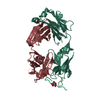


 PDBj
PDBj


 Assembly
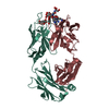

 Mass: 192.124 Da / Num. of mol.: 2 / Source method: obtained synthetically / Formula: C6H8O7
 Mass: 18.015 Da / Num. of mol.: 276 / Source method: isolated from a natural source / Formula: H2O
 Sample preparation
 / Beamline: X13 / Wavelength: 0.8095 Å
 Processing