 Movie
Movie Controller

[English] 日本語
 Yorodumi
- PDB-1a6t: FAB FRAGMENT OF MAB1-IA MONOCLONAL ANTIBODY TO HUMAN RHINOVIRUS 1... -
+ Open data

- Basic information
| Entry | Database: PDB / ID: 1a6t | ||||||
|---|---|---|---|---|---|---|---|
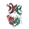
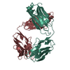
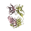
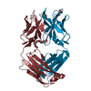
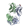
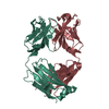
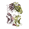
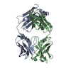
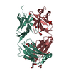
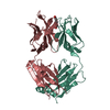
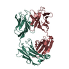
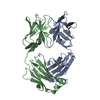
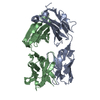
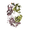
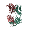
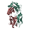
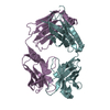
| Title | FAB FRAGMENT OF MAB1-IA MONOCLONAL ANTIBODY TO HUMAN RHINOVIRUS 14 NIM-IA SITE | ||||||
 Components Components |
| ||||||
 Keywords Keywords | IMMUNOGLOBULIN / NEUTRALIZES HUMAN RHINOVIRUS / IGG1 | ||||||
| Function / homology | Immunoglobulins / Immunoglobulin-like / Sandwich / Mainly Beta / : / :  Function and homology information Function and homology information | ||||||
| Biological species |  | ||||||
| Method |  X-RAY DIFFRACTION / MOLECULAR REPLACEMENT / Resolution: 2.7 Å X-RAY DIFFRACTION / MOLECULAR REPLACEMENT / Resolution: 2.7 Å | ||||||
 Authors Authors | Che, Z. / Smith, T.J. | ||||||
 Citation Citation | Journal: J.Virol. / Year: 1998 Title: Antibody-mediated neutralization of human rhinovirus 14 explored by means of cryoelectron microscopy and X-ray crystallography of virus-Fab complexes. Authors: Che, Z. / Olson, N.H. / Leippe, D. / Lee, W.M. / Mosser, A.G. / Rueckert, R.R. / Baker, T.S. / Smith, T.J. | ||||||
| History |
|
- Structure visualization
| Structure viewer | Molecule: MolmilJmol/JSmol |
|---|
- Downloads & links
-Download
| PDBx/mmCIF format | 1a6t.cif.gz | 169.4 KB | Display | PDBx/mmCIF format |
|---|---|---|---|---|
| PDB format | pdb1a6t.ent.gz | 135.3 KB | Display | PDB format |
| PDBx/mmJSON format | 1a6t.json.gz | Tree view | PDBx/mmJSON format | |
| Others |  Other downloads Other downloads |
-Validation report
| Arichive directory | https://data.pdbj.org/pub/pdb/validation_reports/a6/1a6tftp://data.pdbj.org/pub/pdb/validation_reports/a6/1a6t | HTTPS FTP |
|---|
-Related structure data
| Related structure data |  1forS S: Starting model for refinement |
|---|---|
| Similar structure data |


-Links
 PDBj
PDBj


- Assembly
| Deposited unit | 
| ||||||||
|---|---|---|---|---|---|---|---|---|---|
| 1 | 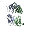
| ||||||||
| 2 | 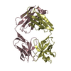
| ||||||||
| Unit cell |
| ||||||||
| Details | THERE ARE TWO FABS IN THE ASYMMETRIC UNIT. THE LIGHT AND HEAVY CHAINS ARE CALLED A AND B, RESPECTIVELY, FOR THE FIRST AND C AND D FOR THE SECOND. |
-Components
| #1: Antibody | Mass: 22697.986 Da / Num. of mol.: 2 / Fragment: FAB FRAGMENT / Source method: isolated from a natural source / Details: MONOCLONAL ANTIBODY / Source: (natural) #2: Antibody | Mass: 23294.027 Da / Num. of mol.: 2 / Fragment: FAB FRAGMENT / Source method: isolated from a natural source / Details: MONOCLONAL ANTIBODY / Source: (natural) Has protein modification | Y | |
|---|
-Experimental details
-Experiment
| Experiment | Method: X-RAY DIFFRACTION / Number of used crystals: 2 |
|---|
- Sample preparation
| Crystal | Density Matthews: 2.76 Å3/Da / Density % sol: 55.43 % | |||||||||||||||||||||||||
|---|---|---|---|---|---|---|---|---|---|---|---|---|---|---|---|---|---|---|---|---|---|---|---|---|---|---|
| Crystal grow | Method: vapor diffusion, sitting drop / pH: 6 Details: SITTING DROP METHOD USING 18-22% PEG8000, 0.1 M SODIUM PHOSPHATE BUFFER (PH 6.0), AND 1% 2-METHYL-2,4-PENTANEDIOL WITH AN FAB1 CONCENTRATION OF 18 MG/ML., vapor diffusion - sitting drop | |||||||||||||||||||||||||
| Crystal grow | *PLUS Method: vapor diffusion, sitting drop | |||||||||||||||||||||||||
| Components of the solutions | *PLUS
|
-Data collection
| Diffraction | Mean temperature: 287 K |
|---|---|
| Diffraction source | Source: ROTATING ANODE / Type: RIGAKU RUH2R / Wavelength: 1.5418 |
| Detector | Type: RIGAKU RAXIS / Detector: IMAGE PLATE / Date: Sep 1, 1996 / Details: MIRRORS |
| Radiation | Monochromator: NI FILTER / Monochromatic (M) / Laue (L): M / Scattering type: x-ray |
| Radiation wavelength | Wavelength: 1.5418 Å / Relative weight: 1 |
| Reflection | Resolution: 2.6→30 Å / Num. obs: 29406 / % possible obs: 91.7 % / Observed criterion σ(I): 2 / Redundancy: 2.4 % / Rmerge(I) obs: 0.042 / Rsym value: 0.042 / Net I/σ(I): 3700 |
| Reflection shell | Resolution: 2.6→2.64 Å / Redundancy: 2 % / Rmerge(I) obs: 0.15 / Mean I/σ(I) obs: 581 / Rsym value: 0.15 / % possible all: 89.2 |
| Reflection shell | *PLUS % possible obs: 89.2 % |
- Processing
| Software |
| ||||||||||||||||||||||||||||||||||||||||||||||||||||||||||||
|---|---|---|---|---|---|---|---|---|---|---|---|---|---|---|---|---|---|---|---|---|---|---|---|---|---|---|---|---|---|---|---|---|---|---|---|---|---|---|---|---|---|---|---|---|---|---|---|---|---|---|---|---|---|---|---|---|---|---|---|---|---|
| Refinement | Method to determine structure: MOLECULAR REPLACEMENT Starting model: PDB ENTRY 1FOR Resolution: 2.7→6 Å / Data cutoff high absF: 10000000 / Data cutoff low absF: 0.001 / Cross valid method: THROUGHOUT / σ(F): 0 / Details: STILL BEING REFINED
| ||||||||||||||||||||||||||||||||||||||||||||||||||||||||||||
| Displacement parameters | Biso mean: 23.2 Å2 | ||||||||||||||||||||||||||||||||||||||||||||||||||||||||||||
| Refinement step | Cycle: LAST / Resolution: 2.7→6 Å
| ||||||||||||||||||||||||||||||||||||||||||||||||||||||||||||
| Refine LS restraints |
| ||||||||||||||||||||||||||||||||||||||||||||||||||||||||||||
| LS refinement shell | Resolution: 2.7→2.81 Å / Total num. of bins used: 6 / % reflection obs: 92 % | ||||||||||||||||||||||||||||||||||||||||||||||||||||||||||||
| Xplor file |
|
