 Movie
Movie Controller


+ Open data

- Basic information
| Entry | Database: PDB / ID: 1ay1 | ||||||
|---|---|---|---|---|---|---|---|
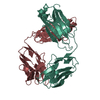
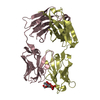
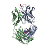
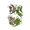
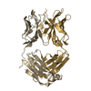
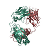
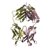
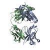
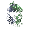
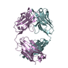
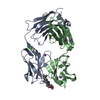
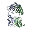
| Title | ANTI TAQ FAB TP7 | ||||||
 Components Components | (TP7 FAB) x 2 | ||||||
 Keywords Keywords | IMMUNOGLOBULIN / ANTIBODY / FAB / ENZYME INHIBITOR / PCR / HOT START | ||||||
| Function / homology | Immunoglobulins / Immunoglobulin-like / Sandwich / Mainly Beta / : / :  Function and homology information Function and homology information | ||||||
| Biological species |  | ||||||
| Method |  X-RAY DIFFRACTION / MOLECULAR REPLACEMENT / Resolution: 2.2 Å X-RAY DIFFRACTION / MOLECULAR REPLACEMENT / Resolution: 2.2 Å | ||||||
 Authors Authors | Murali, R. / Helmer-Citterich, M. / Sharkey, D.J. / Scalice, E.R. / Daiss, J.L. / Sullivan, M.A. / Murthy, H.M.K. | ||||||
 Citation Citation | Journal: Protein Eng. / Year: 1998 Title: Structural studies on an inhibitory antibody against Thermus aquaticus DNA polymerase suggest mode of inhibition. Authors: Murali, R. / Helmer-Citterich, M. / Sharkey, D.J. / Scalice, E.R. / Daiss, J.L. / Sullivan, M.A. / Krishna Murthy, H.M. | ||||||
| History |
|
- Structure visualization
| Structure viewer | Molecule: MolmilJmol/JSmol |
|---|
- Downloads & links
-Download
| PDBx/mmCIF format | 1ay1.cif.gz | 94.6 KB | Display | PDBx/mmCIF format |
|---|---|---|---|---|
| PDB format | pdb1ay1.ent.gz | 71.3 KB | Display | PDB format |
| PDBx/mmJSON format | 1ay1.json.gz | Tree view | PDBx/mmJSON format | |
| Others |  Other downloads Other downloads |
-Validation report
| Arichive directory | https://data.pdbj.org/pub/pdb/validation_reports/ay/1ay1ftp://data.pdbj.org/pub/pdb/validation_reports/ay/1ay1 | HTTPS FTP |
|---|
-Related structure data
| Related structure data |  1hilS S: Starting model for refinement |
|---|---|
| Similar structure data |


-Links
 PDBj
PDBj

- Assembly
| Deposited unit | 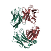
| ||||||||
|---|---|---|---|---|---|---|---|---|---|
| 1 |
| ||||||||
| Unit cell |
|
-Components
| #1: Antibody | Mass: 23082.521 Da / Num. of mol.: 1 / Fragment: FAB / Source method: isolated from a natural source / Source: (natural) |
|---|---|
| #2: Antibody | Mass: 23590.266 Da / Num. of mol.: 1 / Fragment: FAB / Source method: isolated from a natural source / Source: (natural) |
| Has protein modification | Y |
| Sequence details | THE FAB LIGHT CHAIN HAS BEEN ASSIGNED CHAIN INDICATOR *L*. THE FAB HEAVY CHAIN HAS BEEN ASSIGNED ...THE FAB LIGHT CHAIN HAS BEEN ASSIGNED CHAIN INDICATOR *L*. THE FAB HEAVY CHAIN HAS BEEN ASSIGNED CHAIN INDICATOR *H*. FRAGMENT IS NUMBERED ACCORDING TO THE NUMBER CONVENTION |
-Experimental details
-Experiment
| Experiment | Method: X-RAY DIFFRACTION / Number of used crystals: 2 |
|---|
- Sample preparation
| Crystal | Density Matthews: 2.8 Å3/Da / Density % sol: 56 % | |||||||||||||||||||||||||||||||||||
|---|---|---|---|---|---|---|---|---|---|---|---|---|---|---|---|---|---|---|---|---|---|---|---|---|---|---|---|---|---|---|---|---|---|---|---|---|
| Crystal grow | pH: 9 Details: PROTEIN WAS CRYSTALLIZED FROM 16% PEG3350, 0.4% BETA OCTYL GLUCOSIDE, 100MM TRIS, PH 9.0 | |||||||||||||||||||||||||||||||||||
| Crystal grow | *PLUS Temperature: 20 ℃ / Method: vapor diffusion, hanging drop | |||||||||||||||||||||||||||||||||||
| Components of the solutions | *PLUS
|
-Data collection
| Diffraction | Mean temperature: 293 K |
|---|---|
| Diffraction source | Source: ROTATING ANODE / Type: MACSCIENCE / Wavelength: 1.5418 |
| Detector | Type: SIEMENS / Detector: AREA DETECTOR / Date: Oct 1, 1995 |
| Radiation | Monochromator: GRAPHITE(002) / Monochromatic (M) / Laue (L): M / Scattering type: x-ray |
| Radiation wavelength | Wavelength: 1.5418 Å / Relative weight: 1 |
| Reflection | Resolution: 2.2→20 Å / Num. obs: 50786 / % possible obs: 83 % / Observed criterion σ(I): 2 / Redundancy: 3 % / Biso Wilson estimate: 15.5 Å2 / Rmerge(I) obs: 0.078 / Rsym value: 0.056 / Net I/σ(I): 12.8 |
| Reflection shell | Resolution: 2.2→2.3 Å / Redundancy: 2.1 % / Rmerge(I) obs: 0.112 / Mean I/σ(I) obs: 4.8 / Rsym value: 0.068 / % possible all: 68.2 |
| Reflection shell | *PLUS % possible obs: 68.2 % |
- Processing
| Software |
| ||||||||||||||||||||||||||||||||||||||||||||||||||||||||||||||||||||||||||||||||
|---|---|---|---|---|---|---|---|---|---|---|---|---|---|---|---|---|---|---|---|---|---|---|---|---|---|---|---|---|---|---|---|---|---|---|---|---|---|---|---|---|---|---|---|---|---|---|---|---|---|---|---|---|---|---|---|---|---|---|---|---|---|---|---|---|---|---|---|---|---|---|---|---|---|---|---|---|---|---|---|---|---|
| Refinement | Method to determine structure: MOLECULAR REPLACEMENT Starting model: PDB ENTRY 1HIL Resolution: 2.2→7 Å / Data cutoff high absF: 1000000 / Data cutoff low absF: 0.01 / Isotropic thermal model: RESTRAINED / Cross valid method: THROUGHOUT / σ(F): 2 Details: SIDE CHAINS OF RESIDUES 130 - 134 IN THE HEAVY CHAIN ARE DISORDERED AND ARE MODELED TO BE IN THEIR MOST PROBABLE CONFORMATIONS.
| ||||||||||||||||||||||||||||||||||||||||||||||||||||||||||||||||||||||||||||||||
| Displacement parameters | Biso mean: 21.8 Å2 | ||||||||||||||||||||||||||||||||||||||||||||||||||||||||||||||||||||||||||||||||
| Refine analyze | Luzzati coordinate error obs: 0.17 Å / Luzzati d res low obs: 7 Å | ||||||||||||||||||||||||||||||||||||||||||||||||||||||||||||||||||||||||||||||||
| Refinement step | Cycle: LAST / Resolution: 2.2→7 Å
| ||||||||||||||||||||||||||||||||||||||||||||||||||||||||||||||||||||||||||||||||
| Refine LS restraints |
| ||||||||||||||||||||||||||||||||||||||||||||||||||||||||||||||||||||||||||||||||
| LS refinement shell | Resolution: 2.2→2.3 Å / Total num. of bins used: 8
| ||||||||||||||||||||||||||||||||||||||||||||||||||||||||||||||||||||||||||||||||
| Xplor file |
| ||||||||||||||||||||||||||||||||||||||||||||||||||||||||||||||||||||||||||||||||
| Software | *PLUS Name: X-PLOR / Version: 3.1F / Classification: refinement | ||||||||||||||||||||||||||||||||||||||||||||||||||||||||||||||||||||||||||||||||
| Refinement | *PLUS | ||||||||||||||||||||||||||||||||||||||||||||||||||||||||||||||||||||||||||||||||
| Solvent computation | *PLUS | ||||||||||||||||||||||||||||||||||||||||||||||||||||||||||||||||||||||||||||||||
| Displacement parameters | *PLUS | ||||||||||||||||||||||||||||||||||||||||||||||||||||||||||||||||||||||||||||||||
| Refine LS restraints | *PLUS
| ||||||||||||||||||||||||||||||||||||||||||||||||||||||||||||||||||||||||||||||||
| LS refinement shell | *PLUS Rfactor obs: 0.239 |
