 Movie
Movie Controller


+ Open data

- Basic information
| Entry | Database: PDB / ID: 3fct | ||||||
|---|---|---|---|---|---|---|---|
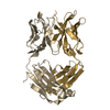
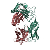
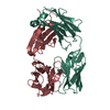
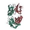
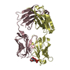
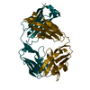
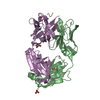
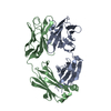
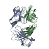
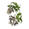
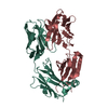
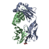
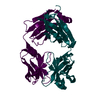
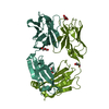
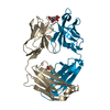
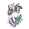
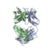
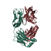
| Title | MATURE METAL CHELATASE CATALYTIC ANTIBODY WITH HAPTEN | ||||||
 Components Components | (PROTEIN (METAL CHELATASE CATALYTIC ...) x 2 | ||||||
 Keywords Keywords | IMMUNE SYSTEM / METAL CHELATASE / CATALYTIC ANTIBODY / FAB FRAGMENT | ||||||
| Function / homology | Immunoglobulins / Immunoglobulin-like / Sandwich / Mainly Beta / : / N-METHYLMESOPORPHYRIN Function and homology information Function and homology information | ||||||
| Biological species |  Homo sapiens (human) Homo sapiens (human) | ||||||
| Method |  X-RAY DIFFRACTION / SYNCHROTRON / MOLECULAR REPLACEMENT / Resolution: 2.4 Å X-RAY DIFFRACTION / SYNCHROTRON / MOLECULAR REPLACEMENT / Resolution: 2.4 Å | ||||||
 Authors Authors | Romesberg, F.E. / Santarsiero, B.D. / Barnes, D. / Yin, J. / Spiller, B. / Schultz, P.G. / Stevens, R.C. | ||||||
 Citation Citation | Journal: Biochemistry / Year: 1998 Title: Structural and kinetic evidence for strain in biological catalysis. Authors: Romesberg, F.E. / Santarsiero, B.D. / Spiller, B. / Yin, J. / Barnes, D. / Schultz, P.G. / Stevens, R.C. | ||||||
| History |
|
- Structure visualization
| Structure viewer | Molecule: MolmilJmol/JSmol |
|---|
- Downloads & links
-Download
| PDBx/mmCIF format | 3fct.cif.gz | 191.2 KB | Display | PDBx/mmCIF format |
|---|---|---|---|---|
| PDB format | pdb3fct.ent.gz | 151.1 KB | Display | PDB format |
| PDBx/mmJSON format | 3fct.json.gz | Tree view | PDBx/mmJSON format | |
| Others |  Other downloads Other downloads |
-Validation report
| Arichive directory | https://data.pdbj.org/pub/pdb/validation_reports/fc/3fctftp://data.pdbj.org/pub/pdb/validation_reports/fc/3fct | HTTPS FTP |
|---|
-Related structure data
| Related structure data | 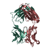 1hklS S: Starting model for refinement |
|---|---|
| Similar structure data |


-Links
 PDBj
PDBj

- Assembly
| Deposited unit | 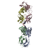
| ||||||||||||||||||||
|---|---|---|---|---|---|---|---|---|---|---|---|---|---|---|---|---|---|---|---|---|---|
| 1 | 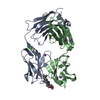
| ||||||||||||||||||||
| 2 | 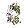
| ||||||||||||||||||||
| Unit cell |
| ||||||||||||||||||||
| Noncrystallographic symmetry (NCS) | NCS oper:
|
-Components
-Antibody , 2 types, 4 molecules ACBD
| #1: Antibody | Mass: 23432.014 Da / Num. of mol.: 2 / Fragment: FAB FRAGMENT Source method: isolated from a genetically manipulated source Details: CHIMERIC FAB FRAGMENT / Source: (gene. exp.) Homo sapiens (human) / Strain: SWISS WEBSTER / Cell: B LYMPHOCYTE / Cell line: 7G12 HYBRIDOMA / Cellular location: PERIPLASM / Organ: SPLEEN / Production host:  #2: Antibody | Mass: 23097.074 Da / Num. of mol.: 2 / Fragment: FAB FRAGMENT Source method: isolated from a genetically manipulated source Details: CHIMERIC FAB FRAGMENT / Source: (gene. exp.) Homo sapiens (human) / Strain: SWISS WEBSTER / Cell: B LYMPHOCYTE / Cell line: 7G12 HYBRIDOMA / Cellular location: PERIPLASM / Organ: SPLEEN / Production host: |
|---|
-Non-polymers , 6 types, 558 molecules 


| #3: Chemical | ChemComp-CD /  Mass: 112.411 Da / Num. of mol.: 1 / Source method: obtained synthetically / Formula: Cd Mass: 112.411 Da / Num. of mol.: 1 / Source method: obtained synthetically / Formula: Cd | ||||
|---|---|---|---|---|---|
| #4: Chemical | ChemComp-CA /  Mass: 40.078 Da / Num. of mol.: 1 / Source method: obtained synthetically / Formula: Ca Mass: 40.078 Da / Num. of mol.: 1 / Source method: obtained synthetically / Formula: Ca | ||||
| #5: Chemical | ChemComp-MG /  Mass: 24.305 Da / Num. of mol.: 1 / Source method: obtained synthetically / Formula: Mg Mass: 24.305 Da / Num. of mol.: 1 / Source method: obtained synthetically / Formula: Mg | ||||
| #6: Chemical | ChemComp-NA /  Mass: 22.990 Da / Num. of mol.: 7 / Source method: obtained synthetically / Formula: Na Mass: 22.990 Da / Num. of mol.: 7 / Source method: obtained synthetically / Formula: Na#7: Chemical |  Mass: 580.716 Da / Num. of mol.: 2 / Source method: obtained synthetically / Formula: C35H40N4O4 Mass: 580.716 Da / Num. of mol.: 2 / Source method: obtained synthetically / Formula: C35H40N4O4#8: Water | ChemComp-HOH / | Mass: 18.015 Da / Num. of mol.: 546 / Source method: isolated from a natural source / Formula: H2O |
-Details
| Has protein modification | Y |
|---|
-Experimental details
-Experiment
| Experiment | Method: X-RAY DIFFRACTION / Number of used crystals: 1 |
|---|
- Sample preparation
| Crystal | Density Matthews: 2.6 Å3/Da / Density % sol: 45 % |
|---|---|
| Crystal grow | Temperature: 293 K / Method: vapor diffusion, hanging drop / pH: 7 Details: PROTEIN WAS CRYSTALLIZED FROM 27% PEG 2000-MME, 200 MM AMMONIUM SULFATE, 100 MM TRIS, PH 7.0, 10MM CADMIUM SULFATE, WITH 10-FOLD EXCESS OF DIASTEREOMERIC N- METHYLMESOPORPHYRIN AT 20C ...Details: PROTEIN WAS CRYSTALLIZED FROM 27% PEG 2000-MME, 200 MM AMMONIUM SULFATE, 100 MM TRIS, PH 7.0, 10MM CADMIUM SULFATE, WITH 10-FOLD EXCESS OF DIASTEREOMERIC N- METHYLMESOPORPHYRIN AT 20C (HANGING DROP)., VAPOR DIFFUSION, HANGING DROP, temperature 293K |
-Data collection
| Diffraction | Mean temperature: 100 K |
|---|---|
| Diffraction source | Source: SYNCHROTRON / Site: NSLS  / Beamline: X12C / Wavelength: 1.1 / Beamline: X12C / Wavelength: 1.1 |
| Detector | Type: MARRESEARCH / Detector: IMAGE PLATE / Date: Oct 15, 1996 / Details: MIRRORS |
| Radiation | Protocol: SINGLE WAVELENGTH / Monochromatic (M) / Laue (L): M / Scattering type: x-ray |
| Radiation wavelength | Wavelength: 1.1 Å / Relative weight: 1 |
| Reflection | Resolution: 2.4→50 Å / % possible obs: 85.9 % / Observed criterion σ(I): 0 / Redundancy: 4 % / Rmerge(I) obs: 0.081 / Net I/σ(I): 18.7 |
- Processing
| Software |
| ||||||||||||||||||||||||||||||||||||||||||||||||||||||||||||||||||||||||||||||||
|---|---|---|---|---|---|---|---|---|---|---|---|---|---|---|---|---|---|---|---|---|---|---|---|---|---|---|---|---|---|---|---|---|---|---|---|---|---|---|---|---|---|---|---|---|---|---|---|---|---|---|---|---|---|---|---|---|---|---|---|---|---|---|---|---|---|---|---|---|---|---|---|---|---|---|---|---|---|---|---|---|---|
| Refinement | Method to determine structure: MOLECULAR REPLACEMENT Starting model: PDB ENTRY 1HKL Resolution: 2.4→50 Å / Isotropic thermal model: RESTRAINED / Cross valid method: THROUGHOUT / σ(F): 2
| ||||||||||||||||||||||||||||||||||||||||||||||||||||||||||||||||||||||||||||||||
| Solvent computation | Solvent model: CNS / Bsol: 45.64 Å2 / ksol: 0.313 e/Å3 | ||||||||||||||||||||||||||||||||||||||||||||||||||||||||||||||||||||||||||||||||
| Refine analyze |
| ||||||||||||||||||||||||||||||||||||||||||||||||||||||||||||||||||||||||||||||||
| Refinement step | Cycle: LAST / Resolution: 2.4→50 Å
| ||||||||||||||||||||||||||||||||||||||||||||||||||||||||||||||||||||||||||||||||
| Refine LS restraints |
| ||||||||||||||||||||||||||||||||||||||||||||||||||||||||||||||||||||||||||||||||
| Xplor file |
|
