| Entry | Database: PDB / ID: 4poz
|
|---|
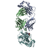
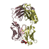
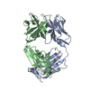
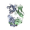
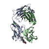
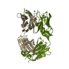
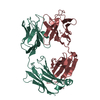
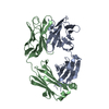
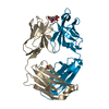
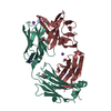
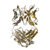
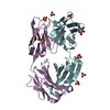
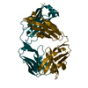
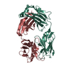
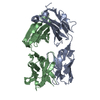
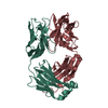
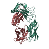
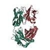
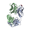
| Title | Fab fragment of Der p 1 specific antibody 10B9 |
|---|
 Components Components | - heavy chain of Fab fragment of 10B9 antibody
- light chain of Fab fragment of 10B9 antibody
|
|---|
 Keywords Keywords | IMMUNE SYSTEM / antibody / allergy |
|---|
| Function / homology | Immunoglobulins / Immunoglobulin-like / Sandwich / Mainly Beta Function and homology information Function and homology information |
|---|
| Biological species |   Mus musculus (house mouse) Mus musculus (house mouse) |
|---|
| Method |  X-RAY DIFFRACTION / SYNCHROTRON / MOLECULAR REPLACEMENT / Resolution: 1.75 Å X-RAY DIFFRACTION / SYNCHROTRON / MOLECULAR REPLACEMENT / Resolution: 1.75 Å |
|---|
 Authors Authors | Osinski, T. / Majorek, K.A. / Pomes, A. / Offermann, L.R. / Osinski, S. / Glesner, J. / Vailes, L.D. / Chapman, M.D. / Minor, W. / Chruszcz, M. |
|---|
 Citation Citation | Journal: J. Immunol. / Year: 2015
Title: Structural Analysis of Der p 1-Antibody Complexes and Comparison with Complexes of Proteins or Peptides with Monoclonal Antibodies.
Authors: Osinski, T. / Pomes, A. / Majorek, K.A. / Glesner, J. / Offermann, L.R. / Vailes, L.D. / Chapman, M.D. / Minor, W. / Chruszcz, M. |
|---|
| History | | Deposition | Feb 26, 2014 | Deposition site: RCSB / Processing site: RCSB |
|---|
| Revision 1.0 | Apr 8, 2015 | Provider: repository / Type: Initial release |
|---|
| Revision 1.1 | Aug 9, 2017 | Group: Database references / Category: citation / citation_author
Item: _citation.country / _citation.journal_abbrev ..._citation.country / _citation.journal_abbrev / _citation.journal_id_CSD / _citation.journal_id_ISSN / _citation.journal_volume / _citation.page_first / _citation.page_last / _citation.pdbx_database_id_DOI / _citation.pdbx_database_id_PubMed / _citation.title / _citation.year |
|---|
| Revision 1.2 | Nov 22, 2017 | Group: Refinement description / Category: software
Item: _software.classification / _software.name / _software.version |
|---|
| Revision 1.3 | Apr 13, 2022 | Group: Database references / Structure summary / Category: audit_author / citation_author / database_2
Item: _audit_author.identifier_ORCID / _citation_author.identifier_ORCID ..._audit_author.identifier_ORCID / _citation_author.identifier_ORCID / _database_2.pdbx_DOI / _database_2.pdbx_database_accession |
|---|
| Revision 1.4 | Sep 20, 2023 | Group: Data collection / Refinement description
Category: chem_comp_atom / chem_comp_bond / pdbx_initial_refinement_model |
|---|
| Revision 1.5 | Nov 6, 2024 | Group: Structure summary / Category: pdbx_entry_details / pdbx_modification_feature |
|---|
|
|---|
 Movie
Movie Controller


 Open data
 Basic information
 Structure visualization
 Downloads & links
 Other downloads
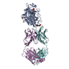


 PDBj
PDBj

 Assembly
 Mass: 18.015 Da / Num. of mol.: 445 / Source method: isolated from a natural source / Formula: H2O
 Sample preparation
 / Beamline: 19-BM / Wavelength: 0.97918 Å
 Processing