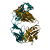
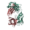
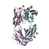
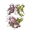
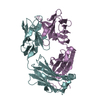
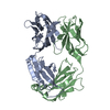
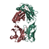
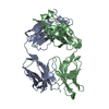
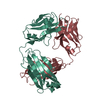
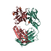
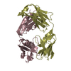
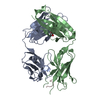
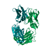
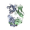
[English] 日本語
Yorodumi
- PDB-4lly: Crystal structure of Pertuzumab Clambda Fab with variable and con... -

+
Open data


ID or keywords:

Loading...

-
Basic information

Entry
Database: PDB / ID: 4lly
TitleCrystal structure of Pertuzumab Clambda Fab with variable and constant domain redesigns (VRD2 and CRD2) at 1.6A
Components
  • light chain Clambda
  • mutated Pertuzumab Fab heavy chain
KeywordsIMMUNE SYSTEM / Fab
Function / homology
Function and homology information


IgD immunoglobulin complex / IgM immunoglobulin complex / IgA immunoglobulin complex / IgE immunoglobulin complex / CD22 mediated BCR regulation / Fc epsilon receptor (FCERI) signaling / Classical antibody-mediated complement activation / Initial triggering of complement / IgG immunoglobulin complex / immunoglobulin complex ...IgD immunoglobulin complex / IgM immunoglobulin complex / IgA immunoglobulin complex / IgE immunoglobulin complex / CD22 mediated BCR regulation / Fc epsilon receptor (FCERI) signaling / Classical antibody-mediated complement activation / Initial triggering of complement / IgG immunoglobulin complex / immunoglobulin complex / immunoglobulin mediated immune response / FCGR activation / Role of phospholipids in phagocytosis / Role of LAT2/NTAL/LAB on calcium mobilization / Scavenging of heme from plasma / antigen binding / FCERI mediated Ca+2 mobilization / FCGR3A-mediated IL10 synthesis / Antigen activates B Cell Receptor (BCR) leading to generation of second messengers / Regulation of Complement cascade / Cell surface interactions at the vascular wall / FCGR3A-mediated phagocytosis / FCERI mediated MAPK activation / B cell receptor signaling pathway / Regulation of actin dynamics for phagocytic cup formation / FCERI mediated NF-kB activation / Immunoregulatory interactions between a Lymphoid and a non-Lymphoid cell / blood microparticle / Potential therapeutics for SARS / adaptive immune response / extracellular space / extracellular exosome / extracellular region / plasma membrane
Similarity search - Function
Immunoglobulin V-Type / Immunoglobulin V-set domain / Immunoglobulin V-set domain / Immunoglobulin subtype / Immunoglobulin / Immunoglobulin/major histocompatibility complex, conserved site / Immunoglobulins and major histocompatibility complex proteins signature. / Immunoglobulin C-Type / Immunoglobulin C1-set / Immunoglobulin C1-set domain ...Immunoglobulin V-Type / Immunoglobulin V-set domain / Immunoglobulin V-set domain / Immunoglobulin subtype / Immunoglobulin / Immunoglobulin/major histocompatibility complex, conserved site / Immunoglobulins and major histocompatibility complex proteins signature. / Immunoglobulin C-Type / Immunoglobulin C1-set / Immunoglobulin C1-set domain / Ig-like domain profile. / Immunoglobulin-like domain / Immunoglobulin-like domain superfamily / Immunoglobulins / Immunoglobulin-like fold / Immunoglobulin-like / Sandwich / Mainly Beta
Similarity search - Domain/homology
Immunoglobulin lambda constant 3 / IgG H chain
Similarity search - Component
Biological speciesHomo sapiens (human)
MethodX-RAY DIFFRACTION / SYNCHROTRON / MOLECULAR REPLACEMENT / Resolution: 1.6 Å
AuthorsPustilnik, A. / Lewis, S.M. / Wu, X. / Sereno, A. / Huang, F. / Guntas, G. / Leaver-Fay, A. / Smith, E.M. / Ho, C. / Hansen-Estruch, C. ...Pustilnik, A. / Lewis, S.M. / Wu, X. / Sereno, A. / Huang, F. / Guntas, G. / Leaver-Fay, A. / Smith, E.M. / Ho, C. / Hansen-Estruch, C. / Chamberlain, A.K. / Truhlar, S.M. / Kuhlman, B. / Demarest, S.J. / Atwell, S.
CitationJournal: Nat.Biotechnol. / Year: 2014
Title: Generation of bispecific IgG antibodies by structure-based design of an orthogonal Fab interface.
Authors: Lewis, S.M. / Wu, X. / Pustilnik, A. / Sereno, A. / Huang, F. / Rick, H.L. / Guntas, G. / Leaver-Fay, A. / Smith, E.M. / Ho, C. / Hansen-Estruch, C. / Chamberlain, A.K. / Truhlar, S.M. / ...Authors: Lewis, S.M. / Wu, X. / Pustilnik, A. / Sereno, A. / Huang, F. / Rick, H.L. / Guntas, G. / Leaver-Fay, A. / Smith, E.M. / Ho, C. / Hansen-Estruch, C. / Chamberlain, A.K. / Truhlar, S.M. / Conner, E.M. / Atwell, S. / Kuhlman, B. / Demarest, S.J.
History
DepositionJul 9, 2013Deposition site: RCSB / Processing site: RCSB
Revision 1.0Jan 29, 2014Provider: repository / Type: Initial release
Revision 1.1Feb 12, 2014Group: Database references
Revision 1.2Mar 26, 2014Group: Database references
Revision 1.3Nov 15, 2017Group: Refinement description / Category: software / Item: _software.name
Revision 2.0Jun 26, 2019Group: Advisory / Data collection ...Advisory / Data collection / Database references / Polymer sequence / Source and taxonomy / Structure summary
Category: entity / entity_poly ...entity / entity_poly / entity_poly_seq / entity_src_gen / pdbx_poly_seq_scheme / pdbx_unobs_or_zero_occ_residues / struct_ref / struct_ref_seq
Item: _entity.formula_weight / _entity_poly.pdbx_seq_one_letter_code ..._entity.formula_weight / _entity_poly.pdbx_seq_one_letter_code / _entity_poly.pdbx_seq_one_letter_code_can / _entity_src_gen.pdbx_beg_seq_num / _entity_src_gen.pdbx_end_seq_num / _entity_src_gen.pdbx_seq_type / _struct_ref.pdbx_align_begin / _struct_ref_seq.db_align_end / _struct_ref_seq.pdbx_auth_seq_align_end / _struct_ref_seq.pdbx_strand_id / _struct_ref_seq.ref_id / _struct_ref_seq.seq_align_end

-
Structure visualization

Structure viewerMolecule:
MolmilJmol/JSmol

Downloads & links

-
Assembly

Deposited unit
A: mutated Pertuzumab Fab heavy chain
B: light chain Clambda
C: mutated Pertuzumab Fab heavy chain
D: light chain Clambda
hetero molecules


Theoretical massNumber of molelcules
Total (without water)94,6729
Polymers94,2794
Non-polymers3935
Water10,467581
1
A: mutated Pertuzumab Fab heavy chain
B: light chain Clambda
hetero molecules


Theoretical massNumber of molelcules
Total (without water)47,3485
Polymers47,1402
Non-polymers2083
Water362
TypeNameSymmetry operationNumber
identity operation1_555x,y,z1
Buried area3790 Å2
ΔGint-32 kcal/mol
Surface area18860 Å2
MethodPISA
2
C: mutated Pertuzumab Fab heavy chain
D: light chain Clambda
hetero molecules


Theoretical massNumber of molelcules
Total (without water)47,3244
Polymers47,1402
Non-polymers1842
Water362
TypeNameSymmetry operationNumber
identity operation1_555x,y,z1
Buried area3600 Å2
ΔGint-27 kcal/mol
Surface area18970 Å2
MethodPISA
Unit cell
Length a, b, c (Å)52.772, 52.847, 89.483
Angle α, β, γ (deg.)76.88, 84.32, 85.53
Int Tables number1
Space group name H-MP1

-
Components

#1: Antibody mutated Pertuzumab Fab heavy chain


Mass: 23973.785 Da / Num. of mol.: 2
Source method: isolated from a genetically manipulated source
Source: (gene. exp.) Homo sapiens (human) / Cell line (production host): HEK293 / Production host: homo sapiens (human) / References: UniProt: S6B291*PLUS
#2: Antibody light chain Clambda


Mass: 23165.801 Da / Num. of mol.: 2
Source method: isolated from a genetically manipulated source
Source: (gene. exp.) Homo sapiens (human) / Cell line (production host): HEK293 / Production host: homo sapiens (human) / References: UniProt: P0DOY3*PLUS
#3: Chemical
ChemComp-GOL / GLYCEROL / GLYCERIN / PROPANE-1,2,3-TRIOL / Glycerol


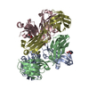
Mass: 92.094 Da / Num. of mol.: 4 / Source method: obtained synthetically / Formula: C3H8O3
#4: Chemical ChemComp-MG / MAGNESIUM ION


Mass: 24.305 Da / Num. of mol.: 1 / Source method: obtained synthetically / Formula: Mg
#5: Water ChemComp-HOH / water / Water


Mass: 18.015 Da / Num. of mol.: 581 / Source method: isolated from a natural source / Formula: H2O

-
Experimental details

-
Experiment

ExperimentMethod: X-RAY DIFFRACTION / Number of used crystals: 1

-
Sample preparation

CrystalDensity Matthews: 1.67 Å3/Da / Density % sol: 26.55 %
Crystal growTemperature: 294 K / Method: vapor diffusion, sitting drop / pH: 5
Details: 100mM Na Citrate tribasic dihydrate pH 5 + 30% Jeffamine ED-2001 , VAPOR DIFFUSION, SITTING DROP, temperature 294K

-
Data collection

Diffraction sourceSource: SYNCHROTRON / Site: APS / Beamline: 31-ID / Wavelength: 0.97931 Å
DetectorType: RAYONIX MX225HE / Detector: CCD / Date: Feb 11, 2013
RadiationMonochromator: Diamond (111) / Protocol: SINGLE WAVELENGTH / Monochromatic (M) / Laue (L): M / Scattering type: x-ray
Radiation wavelengthWavelength: 0.97931 Å / Relative weight: 1
ReflectionResolution: 1.6→43.42 Å / Num. all: 118299 / Num. obs: 118271 / % possible obs: 95.9 % / Observed criterion σ(F): 0 / Observed criterion σ(I): 0
Reflection shellResolution: 1.6→1.69 Å / % possible all: 94.3

-
Processing

Software
NameVersionClassification
MAR345data collection
PHASERphasing
REFMAC5.7.0017refinement
MOSFLMdata reduction
SCALAdata scaling
RefinementMethod to determine structure: MOLECULAR REPLACEMENT / Resolution: 1.6→30 Å / Cor.coef. Fo:Fc: 0.95 / Cor.coef. Fo:Fc free: 0.933 / SU B: 1.746 / SU ML: 0.062 / Cross valid method: THROUGHOUT / ESU R: 0.095 / ESU R Free: 0.095 / Stereochemistry target values: MAXIMUM LIKELIHOOD
RfactorNum. reflection% reflectionSelection details
Rfree0.22929 5989 5.1 %RANDOM
Rwork0.19772 ---
all0.199 118270 --
obs0.19931 112281 95.83 %-
Solvent computationIon probe radii: 0.8 Å / Shrinkage radii: 0.8 Å / VDW probe radii: 1.2 Å / Solvent model: MASK
Displacement parametersBiso mean: 19.076 Å2
Baniso -1Baniso -2Baniso -3
1--0.05 Å2-0.04 Å2-0.11 Å2
2--0.2 Å2-0.48 Å2
3---0.09 Å2
Refinement stepCycle: LAST / Resolution: 1.6→30 Å
ProteinNucleic acidLigandSolventTotal
Num. atoms6432 0 25 581 7038
Refine LS restraints
Refine-IDTypeDev idealDev ideal targetNumber
X-RAY DIFFRACTIONr_bond_refined_d0.0090.026685
X-RAY DIFFRACTIONr_bond_other_d
X-RAY DIFFRACTIONr_angle_refined_deg1.3841.9579138
X-RAY DIFFRACTIONr_angle_other_deg
X-RAY DIFFRACTIONr_dihedral_angle_1_deg6.3875854
X-RAY DIFFRACTIONr_dihedral_angle_2_deg35.03924.333240
X-RAY DIFFRACTIONr_dihedral_angle_3_deg13.092151010
X-RAY DIFFRACTIONr_dihedral_angle_4_deg17.3631518
X-RAY DIFFRACTIONr_chiral_restr0.0920.21043
X-RAY DIFFRACTIONr_gen_planes_refined0.0070.0215010
X-RAY DIFFRACTIONr_gen_planes_other
X-RAY DIFFRACTIONr_nbd_refined
X-RAY DIFFRACTIONr_nbd_other
X-RAY DIFFRACTIONr_nbtor_refined
X-RAY DIFFRACTIONr_nbtor_other
X-RAY DIFFRACTIONr_xyhbond_nbd_refined
X-RAY DIFFRACTIONr_xyhbond_nbd_other
X-RAY DIFFRACTIONr_metal_ion_refined
X-RAY DIFFRACTIONr_metal_ion_other
X-RAY DIFFRACTIONr_symmetry_vdw_refined
X-RAY DIFFRACTIONr_symmetry_vdw_other
X-RAY DIFFRACTIONr_symmetry_hbond_refined
X-RAY DIFFRACTIONr_symmetry_hbond_other
X-RAY DIFFRACTIONr_symmetry_metal_ion_refined
X-RAY DIFFRACTIONr_symmetry_metal_ion_other
X-RAY DIFFRACTIONr_mcbond_it
X-RAY DIFFRACTIONr_mcbond_other
X-RAY DIFFRACTIONr_mcangle_it
X-RAY DIFFRACTIONr_mcangle_other
X-RAY DIFFRACTIONr_scbond_it
X-RAY DIFFRACTIONr_scbond_other
X-RAY DIFFRACTIONr_scangle_it
X-RAY DIFFRACTIONr_scangle_other
X-RAY DIFFRACTIONr_long_range_B_refined
X-RAY DIFFRACTIONr_long_range_B_other
X-RAY DIFFRACTIONr_rigid_bond_restr
X-RAY DIFFRACTIONr_sphericity_free
X-RAY DIFFRACTIONr_sphericity_bonded
LS refinement shellResolution: 1.6→1.641 Å / Total num. of bins used: 20
RfactorNum. reflection% reflection
Rfree0.316 435 -
Rwork0.278 8107 -
obs--93.93 %

+
About Yorodumi

-
News

-
Feb 9, 2022. New format data for meta-information of EMDB entries

New format data for meta-information of EMDB entries

  • Version 3 of the EMDB header file is now the official format.
  • The previous official version 1.9 will be removed from the archive.

Related info.:EMDB header

External links:wwPDB to switch to version 3 of the EMDB data model

-
Aug 12, 2020. Covid-19 info

Covid-19 info

URL: https://pdbj.org/emnavi/covid19.php

New page: Covid-19 featured information page in EM Navigator.

Related info.:Covid-19 info / Mar 5, 2020. Novel coronavirus structure data

+
Mar 5, 2020. Novel coronavirus structure data

Novel coronavirus structure data

Related info.:Yorodumi Speices / Aug 12, 2020. Covid-19 info

External links:COVID-19 featured content - PDBj / Molecule of the Month (242):Coronavirus Proteases

+
Jan 31, 2019. EMDB accession codes are about to change! (news from PDBe EMDB page)

EMDB accession codes are about to change! (news from PDBe EMDB page)

  • The allocation of 4 digits for EMDB accession codes will soon come to an end. Whilst these codes will remain in use, new EMDB accession codes will include an additional digit and will expand incrementally as the available range of codes is exhausted. The current 4-digit format prefixed with “EMD-” (i.e. EMD-XXXX) will advance to a 5-digit format (i.e. EMD-XXXXX), and so on. It is currently estimated that the 4-digit codes will be depleted around Spring 2019, at which point the 5-digit format will come into force.
  • The EM Navigator/Yorodumi systems omit the EMD- prefix.

Related info.:Q: What is EMD? / ID/Accession-code notation in Yorodumi/EM Navigator

External links:EMDB Accession Codes are Changing Soon! / Contact to PDBj

+
Jul 12, 2017. Major update of PDB

Major update of PDB

  • wwPDB released updated PDB data conforming to the new PDBx/mmCIF dictionary.
  • This is a major update changing the version number from 4 to 5, and with Remediation, in which all the entries are updated.
  • In this update, many items about electron microscopy experimental information are reorganized (e.g. em_software).
  • Now, EM Navigator and Yorodumi are based on the updated data.

External links:wwPDB Remediation / Enriched Model Files Conforming to OneDep Data Standards Now Available in the PDB FTP Archive

-
Yorodumi

Thousand views of thousand structures

  • Yorodumi is a browser for structure data from EMDB, PDB, SASBDB, etc.
  • This page is also the successor to EM Navigator detail page, and also detail information page/front-end page for Omokage search.
  • The word "yorodu" (or yorozu) is an old Japanese word meaning "ten thousand". "mi" (miru) is to see.

Related info.:EMDB / PDB / SASBDB / Comparison of 3 databanks / Yorodumi Search / Aug 31, 2016. New EM Navigator & Yorodumi / Yorodumi Papers / Jmol/JSmol / Function and homology information / Changes in new EM Navigator and Yorodumi

Read more