 Movie
Movie Controller


+ Open data

- Basic information
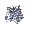
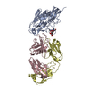
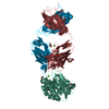
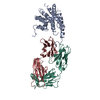
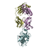
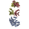
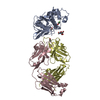
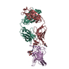
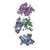
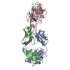
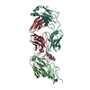
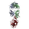
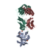
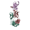
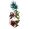
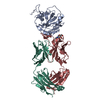
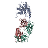
| Entry | Database: PDB / ID: 5vpg | |||||||||
|---|---|---|---|---|---|---|---|---|---|---|
| Title | CRYSTAL STRUCTURE OF DER P 1 COMPLEXED WITH FAB 4C1 | |||||||||
 Components Components |
| |||||||||
 Keywords Keywords | HYDROLASE/IMMUNE SYSTEM / ALLERGEN-ANTIBODY COMPLEX / HYDROLASE-IMMUNE SYSTEM COMPLEX | |||||||||
| Function / homology |  Function and homology information Function and homology information | |||||||||
| Biological species |   Dermatophagoides pteronyssinus (European house dust mite) Dermatophagoides pteronyssinus (European house dust mite) | |||||||||
| Method |  X-RAY DIFFRACTION / SYNCHROTRON / MOLECULAR REPLACEMENT / Resolution: 1.95 Å X-RAY DIFFRACTION / SYNCHROTRON / MOLECULAR REPLACEMENT / Resolution: 1.95 Å | |||||||||
 Authors Authors | Chruszcz, M. / Vailes, L.D. / Chapman, M.D. / Pomes, A. / Minor, W. | |||||||||
 Citation Citation | Journal: J.Biol.Chem. / Year: 2012 Title: Molecular Determinants For Antibody Binding On Group 1 House Dust Mite Allergens. Authors: Chruszcz, M. / Pomes, A. / Glesner, J. / Vailes, L.D. / Osinski, T. / Porebski, P.J. / Majorek, K.A. / Heymann, P.W. / Platts-Mills, T.A. / Minor, W. / Chapman, M.D. | |||||||||
| History |
|
- Structure visualization
| Structure viewer | Molecule: MolmilJmol/JSmol |
|---|
- Downloads & links
-Download
| PDBx/mmCIF format | 5vpg.cif.gz | 291.6 KB | Display | PDBx/mmCIF format |
|---|---|---|---|---|
| PDB format | pdb5vpg.ent.gz | 234.4 KB | Display | PDB format |
| PDBx/mmJSON format | 5vpg.json.gz | Tree view | PDBx/mmJSON format | |
| Others |  Other downloads Other downloads |
-Validation report
| Arichive directory | https://data.pdbj.org/pub/pdb/validation_reports/vp/5vpgftp://data.pdbj.org/pub/pdb/validation_reports/vp/5vpg | HTTPS FTP |
|---|
-Related structure data
| Related structure data |  3rvtC  3rvuC  5vphC  5vplC  3f5vS  3rvv C: citing same article ( S: Starting model for refinement |
|---|---|
| Similar structure data |


-Links
 PDBj
PDBj


- Assembly
| Deposited unit | 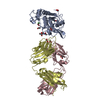
| ||||||||
|---|---|---|---|---|---|---|---|---|---|
| 1 |
| ||||||||
| Unit cell |
|
-Components
-Antibody , 2 types, 2 molecules CD
| #2: Antibody | Mass: 23808.531 Da / Num. of mol.: 1 Source method: isolated from a genetically manipulated source Source: (gene. exp.) |
|---|---|
| #3: Antibody | Mass: 27677.270 Da / Num. of mol.: 1 Source method: isolated from a genetically manipulated source Source: (gene. exp.) |
-Protein / Sugars , 2 types, 2 molecules A
| #1: Protein | Mass: 25014.805 Da / Num. of mol.: 1 / Fragment: UNP RESIDUES 99-320 / Source method: isolated from a natural source Source: (natural) Dermatophagoides pteronyssinus (European house dust mite)References: UniProt: Q3HWZ5 |
|---|---|
| #6: Sugar | ChemComp-NAG /  Type: D-saccharide, beta linking / Mass: 221.208 Da / Num. of mol.: 1 Type: D-saccharide, beta linking / Mass: 221.208 Da / Num. of mol.: 1Source method: isolated from a genetically manipulated source Formula: C8H15NO6 |
-Non-polymers , 3 types, 438 molecules 


| #4: Chemical | ChemComp-CA /  Mass: 40.078 Da / Num. of mol.: 1 / Source method: obtained synthetically / Formula: Ca Mass: 40.078 Da / Num. of mol.: 1 / Source method: obtained synthetically / Formula: Ca | ||
|---|---|---|---|
| #5: Chemical | ChemComp-EDO /  Mass: 62.068 Da / Num. of mol.: 8 / Source method: obtained synthetically / Formula: C2H6O2 Mass: 62.068 Da / Num. of mol.: 8 / Source method: obtained synthetically / Formula: C2H6O2#7: Water | ChemComp-HOH / | Mass: 18.015 Da / Num. of mol.: 429 / Source method: isolated from a natural source / Formula: H2O |
-Details
| Has protein modification | Y |
|---|
-Experimental details
-Experiment
| Experiment | Method: X-RAY DIFFRACTION / Number of used crystals: 1 |
|---|
- Sample preparation
| Crystal | Density Matthews: 2.25 Å3/Da / Density % sol: 45.3 % |
|---|---|
| Crystal grow | Temperature: 293 K / Method: vapor diffusion / pH: 6 Details: 0.1M NA CACODYLATE, 15% W/V PEG4000, PH 6.0, VAPOR DIFFUSION, HANGING DROP, TEMPERATURE 293K PH range: 6 |
-Data collection
| Diffraction | Mean temperature: 100 K |
|---|---|
| Diffraction source | Source: SYNCHROTRON / Site: APS  / Beamline: 19-ID / Wavelength: 0.9794 Å / Beamline: 19-ID / Wavelength: 0.9794 Å |
| Detector | Type: ADSC QUANTUM 315r / Detector: CCD / Date: Mar 21, 2009 / Details: MIRRORS |
| Radiation | Monochromator: SI 111 CHANNEL / Protocol: SINGLE WAVELENGTH / Monochromatic (M) / Laue (L): M / Scattering type: x-ray |
| Radiation wavelength | Wavelength: 0.9794 Å / Relative weight: 1 |
| Reflection | Resolution: 1.95→50 Å / Num. obs: 47483 / % possible obs: 92.9 % / Observed criterion σ(I): -3 / Redundancy: 6.8 % / Rmerge(I) obs: 0.086 / Rsym value: 0.086 / Net I/σ(I): 26 |
| Reflection shell | Resolution: 1.95→1.98 Å / Redundancy: 6.6 % / Rmerge(I) obs: 0.535 / Mean I/σ(I) obs: 3.7 / Rsym value: 0.535 / % possible all: 89.5 |
- Processing
| Software |
| ||||||||||||||||||||||||||||||||||||||||||||||||||||||||||||||||||||||||||||||||||||||||||||||||||||||||||||||||||||||||||||||||||||||||||||||||||||||||||||||||||||||||||||||||||||||
|---|---|---|---|---|---|---|---|---|---|---|---|---|---|---|---|---|---|---|---|---|---|---|---|---|---|---|---|---|---|---|---|---|---|---|---|---|---|---|---|---|---|---|---|---|---|---|---|---|---|---|---|---|---|---|---|---|---|---|---|---|---|---|---|---|---|---|---|---|---|---|---|---|---|---|---|---|---|---|---|---|---|---|---|---|---|---|---|---|---|---|---|---|---|---|---|---|---|---|---|---|---|---|---|---|---|---|---|---|---|---|---|---|---|---|---|---|---|---|---|---|---|---|---|---|---|---|---|---|---|---|---|---|---|---|---|---|---|---|---|---|---|---|---|---|---|---|---|---|---|---|---|---|---|---|---|---|---|---|---|---|---|---|---|---|---|---|---|---|---|---|---|---|---|---|---|---|---|---|---|---|---|---|---|
| Refinement | Method to determine structure: MOLECULAR REPLACEMENT Starting model: PDB ENTRIES 3F5V, 3RVV Resolution: 1.95→45.5 Å / Cor.coef. Fo:Fc: 0.963 / Cor.coef. Fo:Fc free: 0.947 / SU B: 6.438 / SU ML: 0.085 / Cross valid method: THROUGHOUT / ESU R: 0.16 / ESU R Free: 0.144 / Details: HYDROGENS HAVE BEEN ADDED IN THE RIDING POSITIONS
| ||||||||||||||||||||||||||||||||||||||||||||||||||||||||||||||||||||||||||||||||||||||||||||||||||||||||||||||||||||||||||||||||||||||||||||||||||||||||||||||||||||||||||||||||||||||
| Solvent computation | Ion probe radii: 0.8 Å / Shrinkage radii: 0.8 Å / VDW probe radii: 1.4 Å | ||||||||||||||||||||||||||||||||||||||||||||||||||||||||||||||||||||||||||||||||||||||||||||||||||||||||||||||||||||||||||||||||||||||||||||||||||||||||||||||||||||||||||||||||||||||
| Displacement parameters | Biso mean: 28.19 Å2
| ||||||||||||||||||||||||||||||||||||||||||||||||||||||||||||||||||||||||||||||||||||||||||||||||||||||||||||||||||||||||||||||||||||||||||||||||||||||||||||||||||||||||||||||||||||||
| Refinement step | Cycle: LAST / Resolution: 1.95→45.5 Å
| ||||||||||||||||||||||||||||||||||||||||||||||||||||||||||||||||||||||||||||||||||||||||||||||||||||||||||||||||||||||||||||||||||||||||||||||||||||||||||||||||||||||||||||||||||||||
| Refine LS restraints |
|