 Movie
Movie Controller


+ Open data

- Basic information
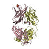
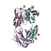
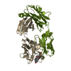
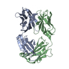
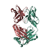
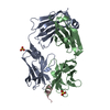
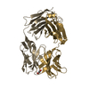
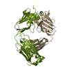
| Entry | Database: PDB / ID: 1kfa | ||||||
|---|---|---|---|---|---|---|---|
| Title | Crystal structure of Fab fragment complexed with gibberellin A4 | ||||||
 Components Components |
| ||||||
 Keywords Keywords | IMMUNE SYSTEM / Immunoglobuiln fold | ||||||
| Function / homology | Immunoglobulins / Immunoglobulin-like / Sandwich / Mainly Beta / GIBBERELLIN A4 Function and homology information Function and homology information | ||||||
| Biological species |  | ||||||
| Method |  X-RAY DIFFRACTION / SYNCHROTRON / MOLECULAR REPLACEMENT / Resolution: 2.8 Å X-RAY DIFFRACTION / SYNCHROTRON / MOLECULAR REPLACEMENT / Resolution: 2.8 Å | ||||||
 Authors Authors | Murata, T. / Fushinobu, S. / Nakajima, M. / Asami, O. / Sassa, T. / Wakagi, T. / Yamaguchi, I. | ||||||
 Citation Citation | Journal: Biochem.Biophys.Res.Commun. / Year: 2002 Title: Crystal structure of the liganded anti-gibberellin A(4) antibody 4-B8(8)/E9 Fab fragment. Authors: Murata, T. / Fushinobu, S. / Nakajima, M. / Asami, O. / Sassa, T. / Wakagi, T. / Yamaguchi, I. #1: Journal: PLANT CELL.PHYSIOL. / Year: 1991Title: Monoclonal antibodies specific for non-derivatized gibberellins I Authors: Nakajima, M. / Yamaguchi, I. / Nagatani, A. / Kizawa, S. / Murofushi, N. / Furuya, N. / Takahashi, N. | ||||||
| History |
|
- Structure visualization
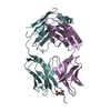
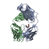
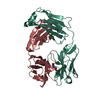
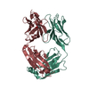
| Structure viewer | Molecule: MolmilJmol/JSmol |
|---|
- Downloads & links
-Download
| PDBx/mmCIF format | 1kfa.cif.gz | 162.7 KB | Display | PDBx/mmCIF format |
|---|---|---|---|---|
| PDB format | pdb1kfa.ent.gz | 131.7 KB | Display | PDB format |
| PDBx/mmJSON format | 1kfa.json.gz | Tree view | PDBx/mmJSON format | |
| Others |  Other downloads Other downloads |
-Validation report
| Arichive directory | https://data.pdbj.org/pub/pdb/validation_reports/kf/1kfaftp://data.pdbj.org/pub/pdb/validation_reports/kf/1kfa | HTTPS FTP |
|---|
-Related structure data
| Related structure data | 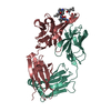 1ikfS S: Starting model for refinement |
|---|---|
| Similar structure data |


-Links
 PDBj
PDBj

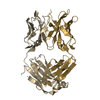
- Assembly
| Deposited unit | 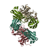
| ||||||||
|---|---|---|---|---|---|---|---|---|---|
| 1 | 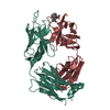
| ||||||||
| 2 | 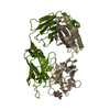
| ||||||||
| 3 | 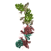
| ||||||||
| Unit cell |
|
-Components
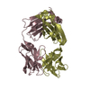
| #1: Antibody | Mass: 23754.285 Da / Num. of mol.: 2 / Fragment: residues 1-217 Source method: isolated from a genetically manipulated source Source: (gene. exp.) #2: Antibody | Mass: 23760.887 Da / Num. of mol.: 2 / Fragment: Variable domain, constant domain 1 Source method: isolated from a genetically manipulated source Source: (gene. exp.) #3: Chemical | 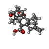  Mass: 332.391 Da / Num. of mol.: 2 / Source method: obtained synthetically / Formula: C19H24O5 Mass: 332.391 Da / Num. of mol.: 2 / Source method: obtained synthetically / Formula: C19H24O5#4: Water | ChemComp-HOH / |  Mass: 18.015 Da / Num. of mol.: 118 / Source method: isolated from a natural source / Formula: H2O Mass: 18.015 Da / Num. of mol.: 118 / Source method: isolated from a natural source / Formula: H2OHas protein modification | Y | |
|---|
-Experimental details
-Experiment
| Experiment | Method: X-RAY DIFFRACTION / Number of used crystals: 1 |
|---|
- Sample preparation
| Crystal | Density Matthews: 2.49 Å3/Da / Density % sol: 50.62 % | ||||||||||||||||||||||||||||||
|---|---|---|---|---|---|---|---|---|---|---|---|---|---|---|---|---|---|---|---|---|---|---|---|---|---|---|---|---|---|---|---|
| Crystal grow | Temperature: 303 K / Method: vapor diffusion, sitting drop / pH: 6.5 Details: PEG 3350, pH 6.5, VAPOR DIFFUSION, SITTING DROP, temperature 303K | ||||||||||||||||||||||||||||||
| Crystal grow | *PLUS Temperature: 30 ℃ | ||||||||||||||||||||||||||||||
| Components of the solutions | *PLUS
|
-Data collection
| Diffraction | Mean temperature: 100 K |
|---|---|
| Diffraction source | Source: SYNCHROTRON / Site: Photon Factory  / Beamline: BL-18B / Wavelength: 1 Å / Beamline: BL-18B / Wavelength: 1 Å |
| Detector | Type: ADSC QUANTUM 4 / Detector: CCD / Date: Oct 19, 2000 |
| Radiation | Monochromator: Si 111 CHANNEL / Protocol: SINGLE WAVELENGTH / Monochromatic (M) / Laue (L): M / Scattering type: x-ray |
| Radiation wavelength | Wavelength: 1 Å / Relative weight: 1 |
| Reflection | Resolution: 2.7→39.62 Å / Num. all: 402565 / Num. obs: 402565 / % possible obs: 100 % / Observed criterion σ(F): 0 / Observed criterion σ(I): 0 / Redundancy: 7.4 % / Biso Wilson estimate: 16.6 Å2 / Rmerge(I) obs: 0.082 / Rsym value: 0.082 / Net I/σ(I): 7.3 |
| Reflection shell | Resolution: 2.7→2.85 Å / Redundancy: 7.5 % / Rmerge(I) obs: 0.321 / Mean I/σ(I) obs: 2.3 / Num. unique all: 3799 / Rsym value: 0.321 / % possible all: 99.7 |
| Reflection | *PLUS Num. obs: 26460 / % possible obs: 100 % / Num. measured all: 402565 |
| Reflection shell | *PLUS Highest resolution: 2.7 Å / % possible obs: 100 % |
- Processing
| Software |
| |||||||||||||||||||||||||
|---|---|---|---|---|---|---|---|---|---|---|---|---|---|---|---|---|---|---|---|---|---|---|---|---|---|---|
| Refinement | Method to determine structure: MOLECULAR REPLACEMENT Starting model: PDB entry 1ikf Resolution: 2.8→39.62 Å / Rfactor Rfree error: 0.009 / Data cutoff high absF: 1685086.35 / Data cutoff low absF: 0 / Isotropic thermal model: Anisotropic / Cross valid method: THROUGHOUT / σ(F): 0 / Stereochemistry target values: Engh & Huber
| |||||||||||||||||||||||||
| Solvent computation | Solvent model: FLAT MODEL / Bsol: 57.0048 Å2 / ksol: 0.373888 e/Å3 | |||||||||||||||||||||||||
| Displacement parameters | Biso mean: 40.7 Å2
| |||||||||||||||||||||||||
| Refine analyze |
| |||||||||||||||||||||||||
| Refinement step | Cycle: LAST / Resolution: 2.8→39.62 Å
| |||||||||||||||||||||||||
| Refine LS restraints |
| |||||||||||||||||||||||||
| LS refinement shell | Resolution: 2.8→2.98 Å / Rfactor Rfree error: 0.025 / Total num. of bins used: 6
| |||||||||||||||||||||||||
| Xplor file |
| |||||||||||||||||||||||||
| Refinement | *PLUS Highest resolution: 2.8 Å / Num. reflection obs: 22549 / % reflection Rfree: 5 % / Rfactor Rfree: 0.296 / Rfactor Rwork: 0.234 | |||||||||||||||||||||||||
| Solvent computation | *PLUS | |||||||||||||||||||||||||
| Displacement parameters | *PLUS | |||||||||||||||||||||||||
| Refine LS restraints | *PLUS
|
