 Movie
Movie Controller

[English] 日本語
 Yorodumi
- PDB-3fo0: Crystal structure of hapten complex of catalytic elimination anti... -
+ Open data

- Basic information
| Entry | Database: PDB / ID: 3fo0 | ||||||
|---|---|---|---|---|---|---|---|
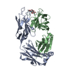
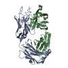
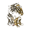
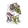
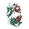
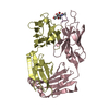
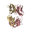
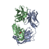
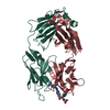
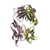
| Title | Crystal structure of hapten complex of catalytic elimination antibody 13G5 (wild-type) | ||||||
 Components Components |
| ||||||
 Keywords Keywords | IMMUNE SYSTEM / IMMUNOGLOBULIN / CATALYTIC ANTIBODY / CHIMERIC FAB / HAPTEN COMPLEX / ACID BASE CATALYSIS / PROTON TRANSFER / Immunoglobulin domain | ||||||
| Function / homology | Immunoglobulins / Immunoglobulin-like / Sandwich / Mainly Beta / Chem-BZH Function and homology information Function and homology information | ||||||
| Biological species |   Homo sapiens (human) Homo sapiens (human) | ||||||
| Method |  X-RAY DIFFRACTION / SYNCHROTRON / MOLECULAR REPLACEMENT / Resolution: 2.5 Å X-RAY DIFFRACTION / SYNCHROTRON / MOLECULAR REPLACEMENT / Resolution: 2.5 Å | ||||||
 Authors Authors | Debler, E.W. / Wilson, I.A. | ||||||
 Citation Citation | Journal: Proc.Natl.Acad.Sci.USA / Year: 2009 Title: An aspartate and a water molecule mediate efficient acid-base catalysis in a tailored antibody pocket. Authors: Debler, E.W. / Muller, R. / Hilvert, D. / Wilson, I.A. | ||||||
| History |
|
- Structure visualization
| Structure viewer | Molecule: MolmilJmol/JSmol |
|---|
- Downloads & links
-Download
| PDBx/mmCIF format | 3fo0.cif.gz | 96.8 KB | Display | PDBx/mmCIF format |
|---|---|---|---|---|
| PDB format | pdb3fo0.ent.gz | 72.2 KB | Display | PDB format |
| PDBx/mmJSON format | 3fo0.json.gz | Tree view | PDBx/mmJSON format | |
| Others |  Other downloads Other downloads |
-Validation report
| Arichive directory | https://data.pdbj.org/pub/pdb/validation_reports/fo/3fo0ftp://data.pdbj.org/pub/pdb/validation_reports/fo/3fo0 | HTTPS FTP |
|---|
-Related structure data
| Related structure data | 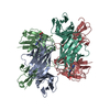 3fo1C 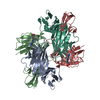 3fo2C  2gjzS C: citing same article ( S: Starting model for refinement |
|---|---|
| Similar structure data |


-Links
 PDBj
PDBj

- Assembly
| Deposited unit | 
| ||||||||
|---|---|---|---|---|---|---|---|---|---|
| 1 |
| ||||||||
| Unit cell |
|
-Components
| #1: Antibody | Mass: 24002.795 Da / Num. of mol.: 1 Source method: isolated from a genetically manipulated source Details: The variable domain (residues 1-107) is from murine source and the constant domain (residues 108-214) is from human source Source: (gene. exp.) Mus musculus, Homo sapiens / Plasmid: p4xH-M13 / Production host:  |
|---|---|
| #2: Antibody | Mass: 24296.205 Da / Num. of mol.: 1 Source method: isolated from a genetically manipulated source Details: The variable domain (residues 1-113) is from murine source and the constant domain (residues 114-235) is from human source Source: (gene. exp.) Mus musculus, Homo sapiens / Plasmid: p4xH-M13 / Production host: |
| #3: Chemical | ChemComp-BZH /   Mass: 262.265 Da / Num. of mol.: 1 / Source method: obtained synthetically / Formula: C12H14N4O3 Mass: 262.265 Da / Num. of mol.: 1 / Source method: obtained synthetically / Formula: C12H14N4O3 |
| #4: Chemical | ChemComp-GOL /   Mass: 92.094 Da / Num. of mol.: 1 / Source method: obtained synthetically / Formula: C3H8O3 Mass: 92.094 Da / Num. of mol.: 1 / Source method: obtained synthetically / Formula: C3H8O3 |
| #5: Water | ChemComp-HOH /  Mass: 18.015 Da / Num. of mol.: 49 / Source method: isolated from a natural source / Formula: H2O Mass: 18.015 Da / Num. of mol.: 49 / Source method: isolated from a natural source / Formula: H2O |
| Has protein modification | Y |
| Sequence details | THE SEQUENCES OF THE FAB COMPLEXES ARE NOT AVAILABLE IN ANY SEQUENCE DATABASES. |
-Experimental details
-Experiment
| Experiment | Method: X-RAY DIFFRACTION / Number of used crystals: 1 |
|---|
- Sample preparation
| Crystal | Density Matthews: 2.25 Å3/Da / Density % sol: 45.3 % |
|---|---|
| Crystal grow | Temperature: 298 K / Method: vapor diffusion, sitting drop / pH: 8.5 Details: 25% PEG 3350, 0.1M Tris-HCl, pH 8.5, VAPOR DIFFUSION, SITTING DROP, temperature 298K |
-Data collection
| Diffraction | Mean temperature: 100 K |
|---|---|
| Diffraction source | Source: SYNCHROTRON / Site: SSRL  / Beamline: BL9-2 / Wavelength: 0.9795 Å / Beamline: BL9-2 / Wavelength: 0.9795 Å |
| Detector | Type: MARMOSAIC 325 mm CCD / Detector: CCD / Date: Aug 4, 2006 / Details: FLAT COLLIMATING MIRROR, TOROID FOCUSING MIRROR |
| Radiation | Monochromator: DOUBLE CRYSTAL Si(111) / Protocol: SINGLE WAVELENGTH / Monochromatic (M) / Laue (L): M / Scattering type: x-ray |
| Radiation wavelength | Wavelength: 0.9795 Å / Relative weight: 1 |
| Reflection | Resolution: 2.5→50 Å / Num. obs: 15696 / % possible obs: 99.7 % / Observed criterion σ(I): -3 / Redundancy: 6.6 % / Rsym value: 0.076 / Net I/σ(I): 36.3 |
| Reflection shell | Resolution: 2.5→2.56 Å / Redundancy: 6.2 % / Mean I/σ(I) obs: 3.8 / Rsym value: 0.552 / % possible all: 99.2 |
- Processing
| Software |
| ||||||||||||||||||||||||||||||||||||||||||||||||||||||||||||||||||||||||||||||||||||||||||||||||||||||||||||||||||||||||||||||||||||||||||||||||||||||||||||||||||||||||||
|---|---|---|---|---|---|---|---|---|---|---|---|---|---|---|---|---|---|---|---|---|---|---|---|---|---|---|---|---|---|---|---|---|---|---|---|---|---|---|---|---|---|---|---|---|---|---|---|---|---|---|---|---|---|---|---|---|---|---|---|---|---|---|---|---|---|---|---|---|---|---|---|---|---|---|---|---|---|---|---|---|---|---|---|---|---|---|---|---|---|---|---|---|---|---|---|---|---|---|---|---|---|---|---|---|---|---|---|---|---|---|---|---|---|---|---|---|---|---|---|---|---|---|---|---|---|---|---|---|---|---|---|---|---|---|---|---|---|---|---|---|---|---|---|---|---|---|---|---|---|---|---|---|---|---|---|---|---|---|---|---|---|---|---|---|---|---|---|---|---|---|---|
| Refinement | Method to determine structure: MOLECULAR REPLACEMENT Starting model: PDB entry 2GJZ Resolution: 2.5→32.39 Å / Cor.coef. Fo:Fc: 0.941 / Cor.coef. Fo:Fc free: 0.915 / SU B: 25.291 / SU ML: 0.265 / TLS residual ADP flag: LIKELY RESIDUAL / Cross valid method: THROUGHOUT / ESU R: 0.856 / ESU R Free: 0.335 / Stereochemistry target values: MAXIMUM LIKELIHOOD / Details: HYDROGENS HAVE BEEN ADDED IN THE RIDING POSITIONS
| ||||||||||||||||||||||||||||||||||||||||||||||||||||||||||||||||||||||||||||||||||||||||||||||||||||||||||||||||||||||||||||||||||||||||||||||||||||||||||||||||||||||||||
| Solvent computation | Ion probe radii: 0.8 Å / Shrinkage radii: 0.8 Å / VDW probe radii: 1.2 Å / Solvent model: BABINET MODEL WITH MASK | ||||||||||||||||||||||||||||||||||||||||||||||||||||||||||||||||||||||||||||||||||||||||||||||||||||||||||||||||||||||||||||||||||||||||||||||||||||||||||||||||||||||||||
| Displacement parameters | Biso mean: 55.185 Å2
| ||||||||||||||||||||||||||||||||||||||||||||||||||||||||||||||||||||||||||||||||||||||||||||||||||||||||||||||||||||||||||||||||||||||||||||||||||||||||||||||||||||||||||
| Refinement step | Cycle: LAST / Resolution: 2.5→32.39 Å
| ||||||||||||||||||||||||||||||||||||||||||||||||||||||||||||||||||||||||||||||||||||||||||||||||||||||||||||||||||||||||||||||||||||||||||||||||||||||||||||||||||||||||||
| Refine LS restraints |
| ||||||||||||||||||||||||||||||||||||||||||||||||||||||||||||||||||||||||||||||||||||||||||||||||||||||||||||||||||||||||||||||||||||||||||||||||||||||||||||||||||||||||||
| LS refinement shell | Resolution: 2.5→2.565 Å / Total num. of bins used: 20
| ||||||||||||||||||||||||||||||||||||||||||||||||||||||||||||||||||||||||||||||||||||||||||||||||||||||||||||||||||||||||||||||||||||||||||||||||||||||||||||||||||||||||||
| Refinement TLS params. | Method: refined / Refine-ID: X-RAY DIFFRACTION
| ||||||||||||||||||||||||||||||||||||||||||||||||||||||||||||||||||||||||||||||||||||||||||||||||||||||||||||||||||||||||||||||||||||||||||||||||||||||||||||||||||||||||||
| Refinement TLS group |
|
