 Movie
Movie Controller

[English] 日本語
 Yorodumi
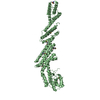
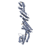
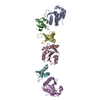
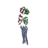
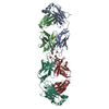
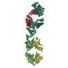
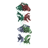
- PDB-1e6j: Crystal structure of HIV-1 capsid protein (p24) in complex with F... -
+ Open data

- Basic information
| Entry | Database: PDB / ID: 1e6j | ||||||
|---|---|---|---|---|---|---|---|
| Title | Crystal structure of HIV-1 capsid protein (p24) in complex with Fab13B5 | ||||||
 Components Components |
| ||||||
 Keywords Keywords | VIRAL PROTEIN / HIV CAPSID PROTEIN (P24) / P24 / FAB / HIV-1 / VIRUS ASSEMBLY / CAPSID / CA / ANTIGEN / ANTIBODY / PROTEIN-PROTEIN INTERACTIONS | ||||||
| Function / homology |  Function and homology information Function and homology informationSynthesis And Processing Of GAG, GAGPOL Polyproteins / host cellular component / host cell nuclear membrane / Integration of viral DNA into host genomic DNA / Autointegration results in viral DNA circles / Minus-strand DNA synthesis / Plus-strand DNA synthesis / Uncoating of the HIV Virion / 2-LTR circle formation / Vpr-mediated nuclear import of PICs ...Synthesis And Processing Of GAG, GAGPOL Polyproteins / host cellular component / host cell nuclear membrane / Integration of viral DNA into host genomic DNA / Autointegration results in viral DNA circles / Minus-strand DNA synthesis / Plus-strand DNA synthesis / Uncoating of the HIV Virion / 2-LTR circle formation / Vpr-mediated nuclear import of PICs / Early Phase of HIV Life Cycle / Integration of provirus / viral budding via host ESCRT complex / APOBEC3G mediated resistance to HIV-1 infection / Binding and entry of HIV virion / immunoglobulin complex / Membrane binding and targetting of GAG proteins / Assembly Of The HIV Virion / Budding and maturation of HIV virion / host multivesicular body / viral nucleocapsid / host cell cytoplasm / adaptive immune response / viral translational frameshifting / host cell nucleus / host cell plasma membrane / virion membrane / structural molecule activity / RNA binding / zinc ion binding / metal ion binding Similarity search - Function | ||||||
| Biological species |  HIV-1 M\:B_HXB2R (virus) HIV-1 M\:B_HXB2R (virus) | ||||||
| Method |  X-RAY DIFFRACTION / SYNCHROTRON / MOLECULAR REPLACEMENT / Resolution: 3 Å X-RAY DIFFRACTION / SYNCHROTRON / MOLECULAR REPLACEMENT / Resolution: 3 Å | ||||||
 Authors Authors | Berthet-Colominas, C. / Monaco, S. / Novelli, A. / Sibai, G. / Mallet, F. / Cusack, S. | ||||||
 Citation Citation | Journal: Structure / Year: 2000 Title: Mutual Conformational Adaptations in Antigen and Antibody Upon Complex Formation between an Fab and HIV-1 Capsid Protein P24 Authors: Monaco-Malbet, S. / Berthet-Colominas, C. / Novelli, A. / Battai, N. / Piga, N. / Cheynet, V. / Mallet, F. / Cusack, S. #1: Journal: Embo J. / Year: 1999 Title: Head-to-Tail Dimers and Interdomain Flexibility Revealed by the Crystal Structure of HIV-1 Capsid Protein (P24) Complexed with a Monoclonal Antibody Fab Authors: Berthet-Colominas, C. / Monaco, S. / Novelli, A. / Sibai, G. / Mallet, F. / Cusack, S. #2: Journal: Protein Expr.Purif. / Year: 1993 Title: Overexpression of HIV-1 Proteins in E. Coli by a Modified Expression Vector and Their One-Step Purification Authors: Cheynet, V. / Verrier, B. / Mallet, F. | ||||||
| History |
|
- Structure visualization
| Structure viewer | Molecule: MolmilJmol/JSmol |
|---|
- Downloads & links
-Download
| PDBx/mmCIF format | 1e6j.cif.gz | 136.1 KB | Display | PDBx/mmCIF format |
|---|---|---|---|---|
| PDB format | pdb1e6j.ent.gz | 106.2 KB | Display | PDB format |
| PDBx/mmJSON format | 1e6j.json.gz | Tree view | PDBx/mmJSON format | |
| Others |  Other downloads Other downloads |
-Validation report
| Arichive directory | https://data.pdbj.org/pub/pdb/validation_reports/e6/1e6jftp://data.pdbj.org/pub/pdb/validation_reports/e6/1e6j | HTTPS FTP |
|---|
-Related structure data
| Related structure data |  1e6oC  1afvS  1am3 S: Starting model for refinement C: citing same article ( |
|---|---|
| Similar structure data |


-Links
 PDBj
PDBj


- Assembly
| Deposited unit | 
| ||||||||
|---|---|---|---|---|---|---|---|---|---|
| 1 |
| ||||||||
| Unit cell |
|
-Components
| #1: Antibody | Mass: 23552.301 Da / Num. of mol.: 1 / Fragment: HEAVY CHAIN 1-219 / Source method: isolated from a natural source / Details: OBTAINED BY PAPAIN CLEAVAGE (FAB) / Source: (natural) |
|---|---|
| #2: Antibody | Mass: 23053.477 Da / Num. of mol.: 1 / Fragment: LIGHT CHAIN 1-210 / Source method: isolated from a natural source / Details: OBTAINED BY PAPAIN CLEAVAGE (FAB) / Source: (natural) |
| #3: Protein | Mass: 23421.795 Da / Num. of mol.: 1 / Fragment: GAG POLYPROTEIN RESIDUES 143-352 Source method: isolated from a genetically manipulated source Details: HIS6 TAG AT N-TERM, PRO1 AND ILE2 DELETED, C-TERMINUS MODIFIED Source: (gene. exp.) HIV-1 M\:B_HXB2R (virus) / Description: SEE REFERENCE 2 / Variant: CLONE 12 / Production host:  |
| Has protein modification | Y |
-Experimental details
-Experiment
| Experiment | Method: X-RAY DIFFRACTION / Number of used crystals: 1 |
|---|
- Sample preparation
| Crystal | Density Matthews: 3.07 Å3/Da / Density % sol: 58.6 % Description: PATTERSON CORRELATION ALGORITHM OF XPLOR USED IN CONJUNCTION WITH AMORE |
|---|---|
| Crystal grow | Temperature: 277 K / Method: vapor diffusion, hanging drop / pH: 7.5 Details: PROTEIN AT 7MG/ML IN 7% PEG8000, 0.1M TRIS-HCL PH=7.5, 5MM DTT, 0.5MM K2PTCL4 AT 4C USING THE HANGING DROP SYSTEM, pH 7.50 |
| Crystal grow | *PLUS Method: unknown |
-Data collection
| Diffraction | Mean temperature: 100 K |
|---|---|
| Diffraction source | Source: SYNCHROTRON / Site: ESRF  / Beamline: ID14-3 / Wavelength: 0.945 / Beamline: ID14-3 / Wavelength: 0.945 |
| Detector | Type: MARRESEARCH / Detector: CCD / Date: Jan 15, 1998 |
| Radiation | Protocol: SINGLE WAVELENGTH / Monochromatic (M) / Laue (L): M / Scattering type: x-ray |
| Radiation wavelength | Wavelength: 0.945 Å / Relative weight: 1 |
| Reflection | Resolution: 3→27.7 Å / Num. obs: 14742 / % possible obs: 84.7 % / Redundancy: 2.4 % / Rsym value: 0.094 |
| Reflection shell | Highest resolution: 3 Å / Redundancy: 1.6 % / Rsym value: 0.276 / % possible all: 33.1 |
| Reflection | *PLUS Rmerge(I) obs: 0.094 |
| Reflection shell | *PLUS % possible obs: 33.1 % / Rmerge(I) obs: 0.276 |
- Processing
| Software |
| ||||||||||||||||||||||||||||||||||||||||||||||||||||||||||||
|---|---|---|---|---|---|---|---|---|---|---|---|---|---|---|---|---|---|---|---|---|---|---|---|---|---|---|---|---|---|---|---|---|---|---|---|---|---|---|---|---|---|---|---|---|---|---|---|---|---|---|---|---|---|---|---|---|---|---|---|---|---|
| Refinement | Method to determine structure: MOLECULAR REPLACEMENT Starting model: FAB13B5, 1AFV, 1AM3 Resolution: 3→15 Å / Cross valid method: THROUGHOUT / σ(F): 0 Details: PARTIALLY DISORDERED REGIONS: L152 - L155 AND H134 - H140 MISSING RESIDUES IN P24: HIS TAG, 12 FIRSTS AA, P146, P86 TO P95
| ||||||||||||||||||||||||||||||||||||||||||||||||||||||||||||
| Refinement step | Cycle: LAST / Resolution: 3→15 Å
| ||||||||||||||||||||||||||||||||||||||||||||||||||||||||||||
| Refine LS restraints |
|
