 Movie
Movie Controller

[English] 日本語
 Yorodumi
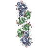
- PDB-4u1g: Plasmodium falciparum reticulocyte-binding protein homologue 5 (P... -
+ Open data

- Basic information
| Entry | Database: PDB / ID: 4u1g | ||||||
|---|---|---|---|---|---|---|---|
| Title | Plasmodium falciparum reticulocyte-binding protein homologue 5 (PfRH5) bound to monoclonal antibody QA1 | ||||||
 Components Components |
| ||||||
 Keywords Keywords | IMMUNE SYSTEM / malaria erythrocyte invasion antibody-mediated inhibition | ||||||
| Function / homology | Rh5 coiled-coil domain / Rh5 coiled-coil domain / Immunoglobulins / Immunoglobulin-like / Sandwich / Mainly Beta / Reticulocyte binding protein 5 Function and homology information Function and homology information | ||||||
| Biological species |   | ||||||
| Method |  X-RAY DIFFRACTION / SYNCHROTRON / MOLECULAR REPLACEMENT / Resolution: 3.1 Å X-RAY DIFFRACTION / SYNCHROTRON / MOLECULAR REPLACEMENT / Resolution: 3.1 Å | ||||||
 Authors Authors | Wright, K.E. / Hjerrild, K.A. / Bartlett, J. / Douglas, A.D. / Jin, J. / Brown, R.E. / Ashfield, R. / Clemmensen, S.B. / de Jongh, W.A. / Draper, S.J. / Higgins, M.K. | ||||||
 Citation Citation | Journal: Nature / Year: 2014 Title: Structure of malaria invasion protein RH5 with erythrocyte basigin and blocking antibodies. Authors: Wright, K.E. / Hjerrild, K.A. / Bartlett, J. / Douglas, A.D. / Jin, J. / Brown, R.E. / Illingworth, J.J. / Ashfield, R. / Clemmensen, S.B. / de Jongh, W.A. / Draper, S.J. / Higgins, M.K. | ||||||
| History |
|
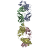
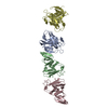
- Structure visualization
| Structure viewer | Molecule: MolmilJmol/JSmol |
|---|
- Downloads & links
-Download
| PDBx/mmCIF format | 4u1g.cif.gz | 596.7 KB | Display | PDBx/mmCIF format |
|---|---|---|---|---|
| PDB format | pdb4u1g.ent.gz | 493.5 KB | Display | PDB format |
| PDBx/mmJSON format | 4u1g.json.gz | Tree view | PDBx/mmJSON format | |
| Others |  Other downloads Other downloads |
-Validation report
| Arichive directory | https://data.pdbj.org/pub/pdb/validation_reports/u1/4u1gftp://data.pdbj.org/pub/pdb/validation_reports/u1/4u1g | HTTPS FTP |
|---|
-Related structure data
| Related structure data | 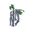 4u0qC  4u0rSC 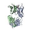 1i7zS 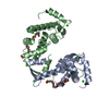 2zn9S S: Starting model for refinement C: citing same article ( |
|---|---|
| Similar structure data |


-Links
 PDBj
PDBj

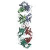
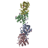
- Assembly
| Deposited unit | 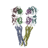
| ||||||||
|---|---|---|---|---|---|---|---|---|---|
| 1 | 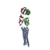
| ||||||||
| 2 | 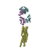
| ||||||||
| Unit cell |
|
-Components
| #1: Protein | Mass: 63128.816 Da / Num. of mol.: 2 / Mutation: yes Source method: isolated from a genetically manipulated source Source: (gene. exp.) Production host:  #2: Antibody | Mass: 27635.135 Da / Num. of mol.: 2 Source method: isolated from a genetically manipulated source Source: (gene. exp.) #3: Antibody | Mass: 26442.330 Da / Num. of mol.: 2 Source method: isolated from a genetically manipulated source Source: (gene. exp.) Has protein modification | Y | |
|---|
-Experimental details
-Experiment
| Experiment | Method: X-RAY DIFFRACTION |
|---|
- Sample preparation
| Crystal | Density Matthews: 2.18 Å3/Da / Density % sol: 43.57 % |
|---|---|
| Crystal grow | Temperature: 291 K / Method: vapor diffusion, sitting drop Details: 0.12 M mix of 1,6-hexanediol, 1-butanol, 1,2-propanediol, 2-propanol, 1,4-butanediol, 1,3-propanediol. 0.1 M MES-imidazole (pH 6.5) 20% (w/v) PEG 550 MM 10% (w/v) PEG 20,000 |
-Data collection
| Diffraction | Mean temperature: 100 K |
|---|---|
| Diffraction source | Source: SYNCHROTRON / Site: Diamond  / Beamline: I04 / Wavelength: 0.97949 Å / Beamline: I04 / Wavelength: 0.97949 Å |
| Detector | Type: PSI PILATUS 6M / Detector: PIXEL / Date: Apr 14, 2014 |
| Radiation | Protocol: SINGLE WAVELENGTH / Monochromatic (M) / Laue (L): M / Scattering type: x-ray |
| Radiation wavelength | Wavelength: 0.97949 Å / Relative weight: 1 |
| Reflection | Resolution: 3.1→47.24 Å / Num. obs: 36313 / % possible obs: 95.5 % / Redundancy: 4.5 % / Biso Wilson estimate: 100.72 Å2 / Rmerge(I) obs: 0.049 / Net I/σ(I): 9.2 |
| Reflection shell | Resolution: 3.1→3.27 Å / Redundancy: 4.6 % / Rmerge(I) obs: 0.38 / Mean I/σ(I) obs: 2.3 / % possible all: 96.3 |
- Processing
| Software |
| |||||||||||||||||||||||||||||||||||||||||||||||||||||||||||||||||||||||||||||||||||||||||||||||||||||||||||||||||||||||||||||||||||||||||||||||||||||||||||||||||||||||||||||||
|---|---|---|---|---|---|---|---|---|---|---|---|---|---|---|---|---|---|---|---|---|---|---|---|---|---|---|---|---|---|---|---|---|---|---|---|---|---|---|---|---|---|---|---|---|---|---|---|---|---|---|---|---|---|---|---|---|---|---|---|---|---|---|---|---|---|---|---|---|---|---|---|---|---|---|---|---|---|---|---|---|---|---|---|---|---|---|---|---|---|---|---|---|---|---|---|---|---|---|---|---|---|---|---|---|---|---|---|---|---|---|---|---|---|---|---|---|---|---|---|---|---|---|---|---|---|---|---|---|---|---|---|---|---|---|---|---|---|---|---|---|---|---|---|---|---|---|---|---|---|---|---|---|---|---|---|---|---|---|---|---|---|---|---|---|---|---|---|---|---|---|---|---|---|---|---|---|
| Refinement | Method to determine structure: MOLECULAR REPLACEMENT Starting model: 4U0R, 1I7Z, 2ZN9 Resolution: 3.1→46.57 Å / Cor.coef. Fo:Fc: 0.8973 / Cor.coef. Fo:Fc free: 0.8572 / Cross valid method: THROUGHOUT / σ(F): 0 / SU Rfree Blow DPI: 0.485
| |||||||||||||||||||||||||||||||||||||||||||||||||||||||||||||||||||||||||||||||||||||||||||||||||||||||||||||||||||||||||||||||||||||||||||||||||||||||||||||||||||||||||||||||
| Displacement parameters | Biso mean: 102.61 Å2
| |||||||||||||||||||||||||||||||||||||||||||||||||||||||||||||||||||||||||||||||||||||||||||||||||||||||||||||||||||||||||||||||||||||||||||||||||||||||||||||||||||||||||||||||
| Refine analyze | Luzzati coordinate error obs: 0.796 Å | |||||||||||||||||||||||||||||||||||||||||||||||||||||||||||||||||||||||||||||||||||||||||||||||||||||||||||||||||||||||||||||||||||||||||||||||||||||||||||||||||||||||||||||||
| Refinement step | Cycle: 1 / Resolution: 3.1→46.57 Å
| |||||||||||||||||||||||||||||||||||||||||||||||||||||||||||||||||||||||||||||||||||||||||||||||||||||||||||||||||||||||||||||||||||||||||||||||||||||||||||||||||||||||||||||||
| Refine LS restraints |
| |||||||||||||||||||||||||||||||||||||||||||||||||||||||||||||||||||||||||||||||||||||||||||||||||||||||||||||||||||||||||||||||||||||||||||||||||||||||||||||||||||||||||||||||
| LS refinement shell | Resolution: 3.1→3.19 Å / Total num. of bins used: 18
| |||||||||||||||||||||||||||||||||||||||||||||||||||||||||||||||||||||||||||||||||||||||||||||||||||||||||||||||||||||||||||||||||||||||||||||||||||||||||||||||||||||||||||||||
| Refinement TLS params. | Method: refined / Refine-ID: X-RAY DIFFRACTION
| |||||||||||||||||||||||||||||||||||||||||||||||||||||||||||||||||||||||||||||||||||||||||||||||||||||||||||||||||||||||||||||||||||||||||||||||||||||||||||||||||||||||||||||||
| Refinement TLS group |
|
