 Movie
Movie Controller


+ Open data

- Basic information
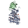
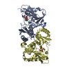
| Entry | Database: PDB / ID: 4m8l | ||||||
|---|---|---|---|---|---|---|---|
| Title | crystal structure of RpiA-R5P complex | ||||||
 Components Components | Ribose-5-phosphate isomerase A | ||||||
 Keywords Keywords | ISOMERASE / Structural Genomics / Center for Structural Genomics of Infectious Diseases / CSGID | ||||||
| Function / homology |  Function and homology information Function and homology informationD-ribose metabolic process / ribose-5-phosphate isomerase / ribose-5-phosphate isomerase activity / pentose-phosphate shunt, non-oxidative branch / cytosol Similarity search - Function | ||||||
| Biological species |  Francisella tularensis (bacteria) Francisella tularensis (bacteria) | ||||||
| Method |  X-RAY DIFFRACTION / SYNCHROTRON / MOLECULAR REPLACEMENT / Resolution: 2.37 Å X-RAY DIFFRACTION / SYNCHROTRON / MOLECULAR REPLACEMENT / Resolution: 2.37 Å | ||||||
 Authors Authors | Rostankowski, R. / Borek, D. / Orlikowska, M. / Nakka, C. / Grimshaw, S. / Otwinowski, Z. / Center for Structural Genomics of Infectious Diseases (CSGID) | ||||||
 Citation Citation | Journal: TO BE PUBLISHED Title: crystal structure of RpiA-R5P complex Authors: Rostankowski, R. / Borek, D. / Orlikowska, M. / Nakka, C. / Grimshaw, S. / Otwinowski, Z. / Center for Structural Genomics of Infectious Diseases (CSGID) | ||||||
| History |
|
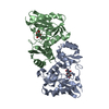
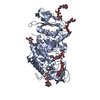
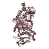
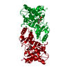
- Structure visualization
| Structure viewer | Molecule: MolmilJmol/JSmol |
|---|
- Downloads & links
-Download
| PDBx/mmCIF format | 4m8l.cif.gz | 187.4 KB | Display | PDBx/mmCIF format |
|---|---|---|---|---|
| PDB format | pdb4m8l.ent.gz | 150.8 KB | Display | PDB format |
| PDBx/mmJSON format | 4m8l.json.gz | Tree view | PDBx/mmJSON format | |
| Others |  Other downloads Other downloads |
-Validation report
| Arichive directory | https://data.pdbj.org/pub/pdb/validation_reports/m8/4m8lftp://data.pdbj.org/pub/pdb/validation_reports/m8/4m8l | HTTPS FTP |
|---|
-Related structure data
| Related structure data |  3kwmS S: Starting model for refinement |
|---|---|
| Similar structure data | |
| Other databases |


-Links
 PDBj
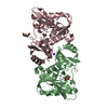
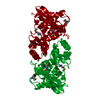
PDBj- Assembly
| Deposited unit | 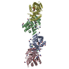
| ||||||||||||||||||||||||||||||||||||||||||||||||||||||||||||||||||||||||||||||||||||||||||||||||||||||||||||||||||||||||||||||||||||||||||||||||||||||
|---|---|---|---|---|---|---|---|---|---|---|---|---|---|---|---|---|---|---|---|---|---|---|---|---|---|---|---|---|---|---|---|---|---|---|---|---|---|---|---|---|---|---|---|---|---|---|---|---|---|---|---|---|---|---|---|---|---|---|---|---|---|---|---|---|---|---|---|---|---|---|---|---|---|---|---|---|---|---|---|---|---|---|---|---|---|---|---|---|---|---|---|---|---|---|---|---|---|---|---|---|---|---|---|---|---|---|---|---|---|---|---|---|---|---|---|---|---|---|---|---|---|---|---|---|---|---|---|---|---|---|---|---|---|---|---|---|---|---|---|---|---|---|---|---|---|---|---|---|---|---|---|
| 1 | 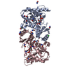
| ||||||||||||||||||||||||||||||||||||||||||||||||||||||||||||||||||||||||||||||||||||||||||||||||||||||||||||||||||||||||||||||||||||||||||||||||||||||
| 2 | 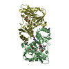
| ||||||||||||||||||||||||||||||||||||||||||||||||||||||||||||||||||||||||||||||||||||||||||||||||||||||||||||||||||||||||||||||||||||||||||||||||||||||
| Unit cell |
| ||||||||||||||||||||||||||||||||||||||||||||||||||||||||||||||||||||||||||||||||||||||||||||||||||||||||||||||||||||||||||||||||||||||||||||||||||||||
| Noncrystallographic symmetry (NCS) | NCS domain:
NCS domain segments: Component-ID: _ / Beg auth comp-ID: ASN / Beg label comp-ID: ASN / Refine code: _
NCS ensembles :
|
-Components
| #1: Protein | Mass: 24494.209 Da / Num. of mol.: 4 Source method: isolated from a genetically manipulated source Source: (gene. exp.) Francisella tularensis (bacteria) / Gene: rpiA, FTW_1255 / Plasmid: pMCSG7 / Production host: #2: Sugar | ChemComp-5RP /   Type: saccharide / Mass: 230.110 Da / Num. of mol.: 4 Type: saccharide / Mass: 230.110 Da / Num. of mol.: 4Source method: isolated from a genetically manipulated source Formula: C5H11O8P #3: Chemical | ChemComp-ARF /   Type: L-peptide NH3 amino terminus / Mass: 45.041 Da / Num. of mol.: 41 / Source method: obtained synthetically / Formula: CH3NO Type: L-peptide NH3 amino terminus / Mass: 45.041 Da / Num. of mol.: 41 / Source method: obtained synthetically / Formula: CH3NO#4: Chemical | ChemComp-CL /   Mass: 35.453 Da / Num. of mol.: 5 / Source method: obtained synthetically / Formula: Cl Mass: 35.453 Da / Num. of mol.: 5 / Source method: obtained synthetically / Formula: Cl#5: Water | ChemComp-HOH / |  Mass: 18.015 Da / Num. of mol.: 292 / Source method: isolated from a natural source / Formula: H2O Mass: 18.015 Da / Num. of mol.: 292 / Source method: isolated from a natural source / Formula: H2O |
|---|
-Experimental details
-Experiment
| Experiment | Method: X-RAY DIFFRACTION / Number of used crystals: 1 |
|---|
- Sample preparation
| Crystal | Density Matthews: 2.36 Å3/Da / Density % sol: 47.91 % |
|---|---|
| Crystal grow | Temperature: 293 K / Method: vapor diffusion, hanging drop / pH: 8.3 Details: 0.22 M magnesium chloride 0.1M Tris pH 6.5, 21% (w/v) PEG 4000, additive: 40% formamide, VAPOR DIFFUSION, HANGING DROP, temperature 293K |
-Data collection
| Diffraction | Mean temperature: 100 K |
|---|---|
| Diffraction source | Source: SYNCHROTRON / Site: APS  / Beamline: 19-ID / Wavelength: 0.97918 Å / Beamline: 19-ID / Wavelength: 0.97918 Å |
| Detector | Type: ADSC QUANTUM 315r / Detector: CCD / Date: Jul 9, 2010 |
| Radiation | Protocol: SINGLE WAVELENGTH / Monochromatic (M) / Laue (L): M / Scattering type: x-ray |
| Radiation wavelength | Wavelength: 0.97918 Å / Relative weight: 1 |
| Reflection | Resolution: 2.37→50 Å / Num. obs: 34431 / % possible obs: 94.5 % / Observed criterion σ(F): -3 / Observed criterion σ(I): 0 / Redundancy: 3.5 % / Rmerge(I) obs: 0.09 / Rsym value: 0.09 / Net I/σ(I): 13.354 |
| Reflection shell | Resolution: 2.4→2.44 Å / Redundancy: 3.1 % / Rmerge(I) obs: 0.328 / Mean I/σ(I) obs: 3.709 / Rsym value: 0.328 / % possible all: 99.9 |
- Processing
| Software |
| ||||||||||||||||||||||||||||||||||||||||||||||||||||||||||||||||||||||||||||||||||||||||||||||||||||||||||||||||||||||||||||||||||||||||||||||||||||||||||||||||||||||||||||||||||||||
|---|---|---|---|---|---|---|---|---|---|---|---|---|---|---|---|---|---|---|---|---|---|---|---|---|---|---|---|---|---|---|---|---|---|---|---|---|---|---|---|---|---|---|---|---|---|---|---|---|---|---|---|---|---|---|---|---|---|---|---|---|---|---|---|---|---|---|---|---|---|---|---|---|---|---|---|---|---|---|---|---|---|---|---|---|---|---|---|---|---|---|---|---|---|---|---|---|---|---|---|---|---|---|---|---|---|---|---|---|---|---|---|---|---|---|---|---|---|---|---|---|---|---|---|---|---|---|---|---|---|---|---|---|---|---|---|---|---|---|---|---|---|---|---|---|---|---|---|---|---|---|---|---|---|---|---|---|---|---|---|---|---|---|---|---|---|---|---|---|---|---|---|---|---|---|---|---|---|---|---|---|---|---|---|
| Refinement | Method to determine structure: MOLECULAR REPLACEMENT Starting model: PDB ENTRY 3KWM Resolution: 2.37→36.75 Å / Cor.coef. Fo:Fc: 0.913 / Cor.coef. Fo:Fc free: 0.89 / SU B: 9.362 / SU ML: 0.227 / Cross valid method: THROUGHOUT / ESU R: 0.79 / ESU R Free: 0.317 / Stereochemistry target values: MAXIMUM LIKELIHOOD / Details: HYDROGENS HAVE BEEN ADDED IN THE RIDING POSITIONS
| ||||||||||||||||||||||||||||||||||||||||||||||||||||||||||||||||||||||||||||||||||||||||||||||||||||||||||||||||||||||||||||||||||||||||||||||||||||||||||||||||||||||||||||||||||||||
| Solvent computation | Ion probe radii: 0.8 Å / Shrinkage radii: 0.8 Å / VDW probe radii: 1.2 Å / Solvent model: BABINET MODEL WITH MASK | ||||||||||||||||||||||||||||||||||||||||||||||||||||||||||||||||||||||||||||||||||||||||||||||||||||||||||||||||||||||||||||||||||||||||||||||||||||||||||||||||||||||||||||||||||||||
| Displacement parameters | Biso mean: 39.183 Å2
| ||||||||||||||||||||||||||||||||||||||||||||||||||||||||||||||||||||||||||||||||||||||||||||||||||||||||||||||||||||||||||||||||||||||||||||||||||||||||||||||||||||||||||||||||||||||
| Refinement step | Cycle: LAST / Resolution: 2.37→36.75 Å
| ||||||||||||||||||||||||||||||||||||||||||||||||||||||||||||||||||||||||||||||||||||||||||||||||||||||||||||||||||||||||||||||||||||||||||||||||||||||||||||||||||||||||||||||||||||||
| Refine LS restraints |
|