 Movie
Movie Controller

[English] 日本語
 Yorodumi
- PDB-4x84: Crystal structure of Ribose-5-phosphate isomerase A from Pseudomo... -
+ Open data

- Basic information
| Entry | Database: PDB / ID: 4x84 | ||||||
|---|---|---|---|---|---|---|---|
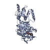
| Title | Crystal structure of Ribose-5-phosphate isomerase A from Pseudomonas aeruginosa | ||||||
 Components Components | Ribose-5-phosphate isomerase A | ||||||
 Keywords Keywords | ISOMERASE / SSGCID / Pseudomonas aeruginosa / Ribose-5-phosphate isomerase A / Phosphoriboisomerase A / PRI / Structural Genomics / Seattle Structural Genomics Center for Infectious Disease | ||||||
| Function / homology |  Function and homology information Function and homology informationD-ribose metabolic process / ribose-5-phosphate isomerase / ribose-5-phosphate isomerase activity / pentose-phosphate shunt, non-oxidative branch / cytosol Similarity search - Function | ||||||
| Biological species |   Pseudomonas aeruginosa (bacteria) Pseudomonas aeruginosa (bacteria) | ||||||
| Method |  X-RAY DIFFRACTION / SYNCHROTRON / MOLECULAR REPLACEMENT / molecular replacement / Resolution: 1.25 Å X-RAY DIFFRACTION / SYNCHROTRON / MOLECULAR REPLACEMENT / molecular replacement / Resolution: 1.25 Å | ||||||
 Authors Authors | Seattle Structural Genomics Center for Infectious Disease (SSGCID) | ||||||
 Citation Citation | Journal: to be published Title: Crystal structure of Ribose-5-phosphate isomerase A from Pseudomonas aeruginosa Authors: Seattle Structural Genomics Center for Infectious Disease (SSGCID) | ||||||
| History |
|
- Structure visualization
| Structure viewer | Molecule: MolmilJmol/JSmol |
|---|
- Downloads & links
-Download
| PDBx/mmCIF format | 4x84.cif.gz | 384.4 KB | Display | PDBx/mmCIF format |
|---|---|---|---|---|
| PDB format | pdb4x84.ent.gz | 310.6 KB | Display | PDB format |
| PDBx/mmJSON format | 4x84.json.gz | Tree view | PDBx/mmJSON format | |
| Others |  Other downloads Other downloads |
-Validation report
| Arichive directory | https://data.pdbj.org/pub/pdb/validation_reports/x8/4x84ftp://data.pdbj.org/pub/pdb/validation_reports/x8/4x84 | HTTPS FTP |
|---|
-Related structure data
| Related structure data |  3enqS S: Starting model for refinement |
|---|---|
| Similar structure data | |
| Other databases |

-Links
 PDBj
PDBj


- Assembly
| Deposited unit | 
| ||||||||
|---|---|---|---|---|---|---|---|---|---|
| 1 | 
| ||||||||
| 2 | 
| ||||||||
| Unit cell |
| ||||||||
| Details | biological unit is a tetramer, there are two biological unics in the asu: chains AD and BC |
-Components
| #1: Protein | Mass: 24780.355 Da / Num. of mol.: 4 / Fragment: PsaeA.00944.a.B1 Source method: isolated from a genetically manipulated source Source: (gene. exp.) Pseudomonas aeruginosa (bacteria) / Strain: ATCC 15692 / PAO1 / 1C / PRS 101 / LMG 12228 / Gene: rpiA, PA0330 / Plasmid: PsaeA.01152.a.B1 / Production host: #2: Chemical | ChemComp-FLC /   Mass: 189.100 Da / Num. of mol.: 4 / Source method: obtained synthetically / Formula: C6H5O7 Mass: 189.100 Da / Num. of mol.: 4 / Source method: obtained synthetically / Formula: C6H5O7#3: Water | ChemComp-HOH / |  Mass: 18.015 Da / Num. of mol.: 1549 / Source method: isolated from a natural source / Formula: H2O Mass: 18.015 Da / Num. of mol.: 1549 / Source method: isolated from a natural source / Formula: H2O |
|---|
-Experimental details
-Experiment
| Experiment | Method: X-RAY DIFFRACTION / Number of used crystals: 1 |
|---|
- Sample preparation
| Crystal | Density Matthews: 2.39 Å3/Da / Density % sol: 48.6 % |
|---|---|
| Crystal grow | Temperature: 290 K / Method: vapor diffusion, sitting drop / pH: 6.5 Details: Moledular Dimensions, Morpheus screen G4: 20mM each Na-Formate; NH4-Acetate; Na3-Citrate; NaKTartrate (racemic); Na-Oxamate; 12.5% each MPD (racemic); PEG 1K; PEG 3350; 100mM Imidazole; MES ...Details: Moledular Dimensions, Morpheus screen G4: 20mM each Na-Formate; NH4-Acetate; Na3-Citrate; NaKTartrate (racemic); Na-Oxamate; 12.5% each MPD (racemic); PEG 1K; PEG 3350; 100mM Imidazole; MES (acid) pH 6.5; PsaeA.00944.a.B1.PS02170 at 29.9mg/ml; direct cryo; tray 258189, puck frv7-1 |
-Data collection
| Diffraction | Mean temperature: 100 K | |||||||||||||||||||||||||||||||||||||||||||||||||||||||||||||||||||||||||||||||||||||||||||||||||||||||||||||||||||||||||||||||||||||||||||||||||||||||||||||||||||||||||||||||||||||||||||||||||||||||||||||||||||||||||||||||||||||||
|---|---|---|---|---|---|---|---|---|---|---|---|---|---|---|---|---|---|---|---|---|---|---|---|---|---|---|---|---|---|---|---|---|---|---|---|---|---|---|---|---|---|---|---|---|---|---|---|---|---|---|---|---|---|---|---|---|---|---|---|---|---|---|---|---|---|---|---|---|---|---|---|---|---|---|---|---|---|---|---|---|---|---|---|---|---|---|---|---|---|---|---|---|---|---|---|---|---|---|---|---|---|---|---|---|---|---|---|---|---|---|---|---|---|---|---|---|---|---|---|---|---|---|---|---|---|---|---|---|---|---|---|---|---|---|---|---|---|---|---|---|---|---|---|---|---|---|---|---|---|---|---|---|---|---|---|---|---|---|---|---|---|---|---|---|---|---|---|---|---|---|---|---|---|---|---|---|---|---|---|---|---|---|---|---|---|---|---|---|---|---|---|---|---|---|---|---|---|---|---|---|---|---|---|---|---|---|---|---|---|---|---|---|---|---|---|---|---|---|---|---|---|---|---|---|---|---|---|---|---|---|---|---|
| Diffraction source | Source: SYNCHROTRON / Site: APS  / Beamline: 21-ID-G / Wavelength: 0.97856 Å / Beamline: 21-ID-G / Wavelength: 0.97856 Å | |||||||||||||||||||||||||||||||||||||||||||||||||||||||||||||||||||||||||||||||||||||||||||||||||||||||||||||||||||||||||||||||||||||||||||||||||||||||||||||||||||||||||||||||||||||||||||||||||||||||||||||||||||||||||||||||||||||||
| Detector | Type: RAYONIX MX-300 / Detector: CCD / Date: Oct 29, 2014 | |||||||||||||||||||||||||||||||||||||||||||||||||||||||||||||||||||||||||||||||||||||||||||||||||||||||||||||||||||||||||||||||||||||||||||||||||||||||||||||||||||||||||||||||||||||||||||||||||||||||||||||||||||||||||||||||||||||||
| Radiation | Monochromator: C(111) / Protocol: SINGLE WAVELENGTH / Monochromatic (M) / Laue (L): M / Scattering type: x-ray | |||||||||||||||||||||||||||||||||||||||||||||||||||||||||||||||||||||||||||||||||||||||||||||||||||||||||||||||||||||||||||||||||||||||||||||||||||||||||||||||||||||||||||||||||||||||||||||||||||||||||||||||||||||||||||||||||||||||
| Radiation wavelength | Wavelength: 0.97856 Å / Relative weight: 1 | |||||||||||||||||||||||||||||||||||||||||||||||||||||||||||||||||||||||||||||||||||||||||||||||||||||||||||||||||||||||||||||||||||||||||||||||||||||||||||||||||||||||||||||||||||||||||||||||||||||||||||||||||||||||||||||||||||||||
| Reflection | Resolution: 1.25→50 Å / Num. all: 262311 / Num. obs: 261250 / % possible obs: 99.6 % / Observed criterion σ(I): -3 / Redundancy: 4.8 % / Biso Wilson estimate: 8.68 Å2 / Rmerge F obs: 0.999 / Rmerge(I) obs: 0.068 / Rrim(I) all: 0.076 / Χ2: 0.95 / Net I/σ(I): 14 / Num. measured all: 1258596 | |||||||||||||||||||||||||||||||||||||||||||||||||||||||||||||||||||||||||||||||||||||||||||||||||||||||||||||||||||||||||||||||||||||||||||||||||||||||||||||||||||||||||||||||||||||||||||||||||||||||||||||||||||||||||||||||||||||||
| Reflection shell | Diffraction-ID: 1 / Rejects: _
|
-Phasing
| Phasing | Method: molecular replacement | |||||||||
|---|---|---|---|---|---|---|---|---|---|---|
| Phasing MR | Model details: Phaser MODE: MR_AUTO
|
- Processing
| Software |
| |||||||||||||||||||||||||||||||||||||||||||||||||||||||||||||||||||||||||||||||||||||||||||||||||||||||||
|---|---|---|---|---|---|---|---|---|---|---|---|---|---|---|---|---|---|---|---|---|---|---|---|---|---|---|---|---|---|---|---|---|---|---|---|---|---|---|---|---|---|---|---|---|---|---|---|---|---|---|---|---|---|---|---|---|---|---|---|---|---|---|---|---|---|---|---|---|---|---|---|---|---|---|---|---|---|---|---|---|---|---|---|---|---|---|---|---|---|---|---|---|---|---|---|---|---|---|---|---|---|---|---|---|---|---|
| Refinement | Method to determine structure: MOLECULAR REPLACEMENT Starting model: PDB entry 3enq Resolution: 1.25→45.649 Å / SU ML: 0.09 / Cross valid method: FREE R-VALUE / σ(F): 1.34 / Phase error: 13.62 / Stereochemistry target values: ML
| |||||||||||||||||||||||||||||||||||||||||||||||||||||||||||||||||||||||||||||||||||||||||||||||||||||||||
| Solvent computation | Shrinkage radii: 0.9 Å / VDW probe radii: 1.11 Å / Solvent model: FLAT BULK SOLVENT MODEL | |||||||||||||||||||||||||||||||||||||||||||||||||||||||||||||||||||||||||||||||||||||||||||||||||||||||||
| Displacement parameters | Biso max: 55.09 Å2 / Biso mean: 14.3851 Å2 / Biso min: 4.78 Å2 | |||||||||||||||||||||||||||||||||||||||||||||||||||||||||||||||||||||||||||||||||||||||||||||||||||||||||
| Refinement step | Cycle: final / Resolution: 1.25→45.649 Å
| |||||||||||||||||||||||||||||||||||||||||||||||||||||||||||||||||||||||||||||||||||||||||||||||||||||||||
| Refine LS restraints |
| |||||||||||||||||||||||||||||||||||||||||||||||||||||||||||||||||||||||||||||||||||||||||||||||||||||||||
| LS refinement shell | Refine-ID: X-RAY DIFFRACTION / Total num. of bins used: 14
|
