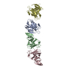
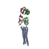
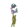
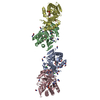
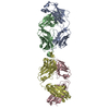
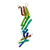
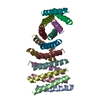
A: Basal-body rod modification protein FlgD B: Basal-body rod modification protein FlgD C: Basal-body rod modification protein FlgD D: Basal-body rod modification protein FlgD
Resolution: 2.75→38.39 Å / Cor.coef. Fo:Fc: 0.931 / Cor.coef. Fo:Fc free: 0.896 / SU B: 16.528 / SU ML: 0.312 / Cross valid method: THROUGHOUT / ESU R Free: 0.416 / Stereochemistry target values: MAXIMUM LIKELIHOOD / Details: HYDROGENS HAVE BEEN ADDED IN THE RIDING POSITIONS
Rfactor
Num. reflection
% reflection
Selection details
Rfree
0.27487
1838
10 %
RANDOM
Rwork
0.22067
-
-
-
obs
0.22599
16537
99.58 %
-
Solvent computation
Ion probe radii: 0.8 Å / Shrinkage radii: 0.8 Å / VDW probe radii: 1.2 Å / Solvent model: MASK
 Movie
Movie Controller

 Yorodumi
 Open data
 Basic information
 Components
 Keywords
 Function and homology information
 Helicobacter pylori (bacteria)
 X-RAY DIFFRACTION /
  Authors
 Italy,
  Croatia, 3items
  Citation
 Structure visualization
 Downloads & links
 Other downloads
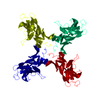


 PDBj
PDBj Assembly
 Mass: 18.015 Da / Num. of mol.: 44 / Source method: isolated from a natural source / Formula: H2O
 Sample preparation
 / Beamline: ID14-4 / Wavelength: 0.95372 Å
 Processing