 Movie
Movie Controller

[English] 日本語
 Yorodumi
- PDB-1rz8: CRYSTAL STRUCTURE OF HUMAN ANTI-HIV-1 GP120-REACTIVE ANTIBODY 17B -
+ Open data

- Basic information
| Entry | Database: PDB / ID: 1rz8 | ||||||
|---|---|---|---|---|---|---|---|
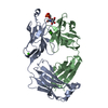
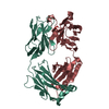
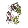
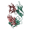
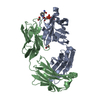
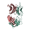
| Title | CRYSTAL STRUCTURE OF HUMAN ANTI-HIV-1 GP120-REACTIVE ANTIBODY 17B | ||||||
 Components Components |
| ||||||
 Keywords Keywords | IMMUNE SYSTEM / HIV-1 / gp120 / CD4i / antibodies / tyrosine sulfation / VH-gene usage | ||||||
| Function / homology | Immunoglobulins / Immunoglobulin-like / Sandwich / Mainly Beta Function and homology information Function and homology information | ||||||
| Biological species |  Homo sapiens (human) Homo sapiens (human) | ||||||
| Method |  X-RAY DIFFRACTION / MOLECULAR REPLACEMENT / Resolution: 2.3 Å X-RAY DIFFRACTION / MOLECULAR REPLACEMENT / Resolution: 2.3 Å | ||||||
 Authors Authors | Huang, C.C. / Venturi, M. / Majeed, S. / Moore, M.J. / Phogat, S. / Zhang, M.-Y. / Dimitrov, D.S. / Hendrickson, W.A. / Robinson, J. / Sodroski, J. ...Huang, C.C. / Venturi, M. / Majeed, S. / Moore, M.J. / Phogat, S. / Zhang, M.-Y. / Dimitrov, D.S. / Hendrickson, W.A. / Robinson, J. / Sodroski, J. / Wyatt, R. / Choe, H. / Farzan, M. / Kwong, P.D. | ||||||
 Citation Citation | Journal: Proc.Natl.Acad.Sci.USA / Year: 2004 Title: Structural basis of tyrosine sulfation and VH-gene usage in antibodies that recognize the HIV type 1 coreceptor-binding site on gp120 Authors: Huang, C.C. / Venturi, M. / Majeed, S. / Moore, M.J. / Phogat, S. / Zhang, M.-Y. / Dimitrov, D.S. / Hendrickson, W.A. / Robinson, J. / Sodroski, J. / Wyatt, R. / Choe, H. / Farzan, M. / Kwong, P.D. | ||||||
| History |
|
- Structure visualization
| Structure viewer | Molecule: MolmilJmol/JSmol |
|---|
- Downloads & links
-Download
| PDBx/mmCIF format | 1rz8.cif.gz | 187.7 KB | Display | PDBx/mmCIF format |
|---|---|---|---|---|
| PDB format | pdb1rz8.ent.gz | 149 KB | Display | PDB format |
| PDBx/mmJSON format | 1rz8.json.gz | Tree view | PDBx/mmJSON format | |
| Others |  Other downloads Other downloads |
-Validation report
| Arichive directory | https://data.pdbj.org/pub/pdb/validation_reports/rz/1rz8ftp://data.pdbj.org/pub/pdb/validation_reports/rz/1rz8 | HTTPS FTP |
|---|
-Related structure data
| Related structure data | 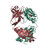 1rz7C 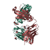 1rzfC 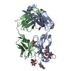 1rzgC 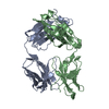 1rziC  1rzjC  1rzkC  1bbjS C: citing same article ( S: Starting model for refinement |
|---|---|
| Similar structure data |


-Links
 PDBj
PDBj


- Assembly
| Deposited unit | 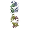
| ||||||||||
|---|---|---|---|---|---|---|---|---|---|---|---|
| 1 | 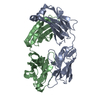
| ||||||||||
| 2 | 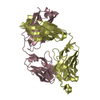
| ||||||||||
| Unit cell |
|
-Components
| #1: Antibody | Mass: 23399.898 Da / Num. of mol.: 2 Source method: isolated from a genetically manipulated source Source: (gene. exp.) Homo sapiens (human) / Genus (production host): LymphocryptovirusCell (production host): IMMORTALIZED B-CELL CLONE FUSED WITH A MURINE B-CELL FUSION PARTNER Production host:  Human herpesvirus 4 (Epstein-Barr virus) Human herpesvirus 4 (Epstein-Barr virus)#2: Antibody | Mass: 24328.205 Da / Num. of mol.: 2 Source method: isolated from a genetically manipulated source Source: (gene. exp.) Homo sapiens (human) / Genus (production host): LymphocryptovirusCell (production host): IMMORTALIZED B-CELL CLONE FUSED WITH A MURINE B-CELL FUSION PARTNER Production host: Human herpesvirus 4 (Epstein-Barr virus)#3: Water | ChemComp-HOH / |  Mass: 18.015 Da / Num. of mol.: 474 / Source method: isolated from a natural source / Formula: H2O Mass: 18.015 Da / Num. of mol.: 474 / Source method: isolated from a natural source / Formula: H2OHas protein modification | Y | |
|---|
-Experimental details
-Experiment
| Experiment | Method: X-RAY DIFFRACTION / Number of used crystals: 1 |
|---|
- Sample preparation
| Crystal | Density Matthews: 3.01 Å3/Da / Density % sol: 59.14 % | ||||||||||||||||||||||||||||||||||||||||||||||||||||||||
|---|---|---|---|---|---|---|---|---|---|---|---|---|---|---|---|---|---|---|---|---|---|---|---|---|---|---|---|---|---|---|---|---|---|---|---|---|---|---|---|---|---|---|---|---|---|---|---|---|---|---|---|---|---|---|---|---|---|
| Crystal grow | Temperature: 293 K / Method: vapor diffusion, hanging drop / pH: 6.5 Details: 50% saturated ammonium sulfate, 10% 2-methyl-2,4-pentanediol, 0.1M imidazole, pH 6.5, VAPOR DIFFUSION, HANGING DROP, temperature 293K | ||||||||||||||||||||||||||||||||||||||||||||||||||||||||
| Crystal grow | *PLUS pH: 7 / Method: vapor diffusion, hanging drop | ||||||||||||||||||||||||||||||||||||||||||||||||||||||||
| Components of the solutions | *PLUS
|
-Data collection
| Diffraction | Mean temperature: 100 K |
|---|---|
| Diffraction source | Source: ROTATING ANODE / Type: RIGAKU / Wavelength: 1.5418 Å |
| Detector | Type: RIGAKU RAXIS / Detector: IMAGE PLATE / Date: Jan 18, 1996 |
| Radiation | Protocol: SINGLE WAVELENGTH / Monochromatic (M) / Laue (L): M / Scattering type: x-ray |
| Radiation wavelength | Wavelength: 1.5418 Å / Relative weight: 1 |
| Reflection | Resolution: 2.2→20 Å / Num. obs: 55222 / Observed criterion σ(I): -3 / Biso Wilson estimate: 19.7 Å2 |
| Reflection shell | Resolution: 2.2→2.28 Å / % possible all: 70.8 |
| Reflection | *PLUS % possible obs: 93.1 % / Num. measured all: 228323 / Rmerge(I) obs: 0.086 |
| Reflection shell | *PLUS % possible obs: 70.8 % |
- Processing
| Software |
| ||||||||||||||||||||||||||||||||||||
|---|---|---|---|---|---|---|---|---|---|---|---|---|---|---|---|---|---|---|---|---|---|---|---|---|---|---|---|---|---|---|---|---|---|---|---|---|---|
| Refinement | Method to determine structure: MOLECULAR REPLACEMENT Starting model: PDB entry 1BBJ Resolution: 2.3→19.99 Å / Rfactor Rfree error: 0.004 / Data cutoff high absF: 200629.33 / Data cutoff low absF: 0 / Isotropic thermal model: RESTRAINED / Cross valid method: THROUGHOUT / σ(F): 0
| ||||||||||||||||||||||||||||||||||||
| Solvent computation | Solvent model: FLAT MODEL / Bsol: 31.7512 Å2 / ksol: 0.340093 e/Å3 | ||||||||||||||||||||||||||||||||||||
| Displacement parameters | Biso mean: 29 Å2
| ||||||||||||||||||||||||||||||||||||
| Refine analyze |
| ||||||||||||||||||||||||||||||||||||
| Refinement step | Cycle: LAST / Resolution: 2.3→19.99 Å
| ||||||||||||||||||||||||||||||||||||
| Refine LS restraints |
| ||||||||||||||||||||||||||||||||||||
| LS refinement shell | Resolution: 2.3→2.44 Å / Rfactor Rfree error: 0.012 / Total num. of bins used: 6
| ||||||||||||||||||||||||||||||||||||
| Xplor file |
| ||||||||||||||||||||||||||||||||||||
| Refinement | *PLUS Highest resolution: 2.3 Å / Lowest resolution: 20 Å / % reflection Rfree: 10 % | ||||||||||||||||||||||||||||||||||||
| Solvent computation | *PLUS | ||||||||||||||||||||||||||||||||||||
| Displacement parameters | *PLUS | ||||||||||||||||||||||||||||||||||||
| Refine LS restraints | *PLUS
|
