 Movie
Movie Controller

[English] 日本語
 Yorodumi
- PDB-6wgk: Fab portion of dupilumab with Crystal Kappa design and intrachain... -
+ Open data

- Basic information
| Entry | Database: PDB / ID: 6wgk | ||||||
|---|---|---|---|---|---|---|---|
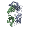
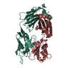
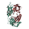
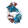
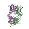
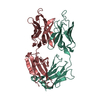
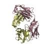
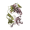
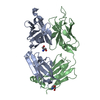
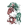
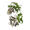
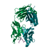
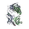
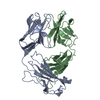
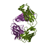
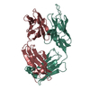
| Title | Fab portion of dupilumab with Crystal Kappa design and intrachain disulfide | ||||||
 Components Components |
| ||||||
 Keywords Keywords | IMMUNE SYSTEM / Dupilumab / hIL4R | ||||||
| Function / homology | Immunoglobulins / Immunoglobulin-like / Sandwich / Mainly Beta Function and homology information Function and homology information | ||||||
| Biological species |  Homo sapiens (human) Homo sapiens (human) | ||||||
| Method |  X-RAY DIFFRACTION / SYNCHROTRON / MOLECULAR REPLACEMENT / Resolution: 1.62 Å X-RAY DIFFRACTION / SYNCHROTRON / MOLECULAR REPLACEMENT / Resolution: 1.62 Å | ||||||
 Authors Authors | Druzina, Z. / Atwell, S. / Pustilnik, A. / Antonysamy, S. / Ho, C. / Lieu, R. / Hendle, J. / Benach, J. / Wang, J. | ||||||
 Citation Citation | Journal: Plos One / Year: 2020 Title: Rapid and robust antibody Fab fragment crystallization utilizing edge-to-edge beta-sheet packing. Authors: Lieu, R. / Antonysamy, S. / Druzina, Z. / Ho, C. / Kang, N.R. / Pustilnik, A. / Wang, J. / Atwell, S. | ||||||
| History |
|
- Structure visualization
| Structure viewer | Molecule: MolmilJmol/JSmol |
|---|
- Downloads & links
-Download
| PDBx/mmCIF format | 6wgk.cif.gz | 344 KB | Display | PDBx/mmCIF format |
|---|---|---|---|---|
| PDB format | pdb6wgk.ent.gz | 277.5 KB | Display | PDB format |
| PDBx/mmJSON format | 6wgk.json.gz | Tree view | PDBx/mmJSON format | |
| Others |  Other downloads Other downloads |
-Validation report
| Arichive directory | https://data.pdbj.org/pub/pdb/validation_reports/wg/6wgkftp://data.pdbj.org/pub/pdb/validation_reports/wg/6wgk | HTTPS FTP |
|---|
-Related structure data
| Related structure data | 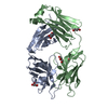 6wg8C 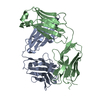 6wgbSC 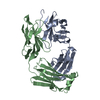 6wgjC  6wglC  6wioC  6wirC S: Starting model for refinement C: citing same article ( |
|---|---|
| Similar structure data |


-Links
 PDBj
PDBj

- Assembly
| Deposited unit | 
| ||||||||
|---|---|---|---|---|---|---|---|---|---|
| 1 | 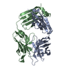
| ||||||||
| 2 | 
| ||||||||
| 3 | 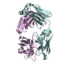
| ||||||||
| 4 | 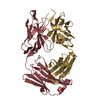
| ||||||||
| Unit cell |
|
-Components
| #1: Antibody | Mass: 25230.133 Da / Num. of mol.: 4 Source method: isolated from a genetically manipulated source Source: (gene. exp.) Homo sapiens (human) / Production host:   Cricetulus griseus (Chinese hamster) Cricetulus griseus (Chinese hamster)#2: Antibody | Mass: 23803.469 Da / Num. of mol.: 4 Source method: isolated from a genetically manipulated source Source: (gene. exp.) Homo sapiens (human) / Production host: Cricetulus griseus (Chinese hamster)#3: Water | ChemComp-HOH / |  Mass: 18.015 Da / Num. of mol.: 791 / Source method: isolated from a natural source / Formula: H2O Mass: 18.015 Da / Num. of mol.: 791 / Source method: isolated from a natural source / Formula: H2OHas protein modification | Y | |
|---|
-Experimental details
-Experiment
| Experiment | Method: X-RAY DIFFRACTION / Number of used crystals: 1 |
|---|
- Sample preparation
| Crystal | Density Matthews: 2.42 Å3/Da / Density % sol: 49.07 % |
|---|---|
| Crystal grow | Temperature: 294 K / Method: vapor diffusion, sitting drop Details: 100mM Tris HCl pH 8.5, 20% PEG MME 2K , 200mM Trimethylamine N-oxide dihydrate |
-Data collection
| Diffraction | Mean temperature: 100 K / Serial crystal experiment: N | ||||||||||||||||||||||||||||||
|---|---|---|---|---|---|---|---|---|---|---|---|---|---|---|---|---|---|---|---|---|---|---|---|---|---|---|---|---|---|---|---|
| Diffraction source | Source: SYNCHROTRON / Site: APS  / Beamline: 31-ID / Wavelength: 0.9793 Å / Beamline: 31-ID / Wavelength: 0.9793 Å | ||||||||||||||||||||||||||||||
| Detector | Type: RAYONIX MX225-HS / Detector: CCD / Date: Feb 5, 2019 | ||||||||||||||||||||||||||||||
| Radiation | Protocol: SINGLE WAVELENGTH / Monochromatic (M) / Laue (L): M / Scattering type: x-ray | ||||||||||||||||||||||||||||||
| Radiation wavelength | Wavelength: 0.9793 Å / Relative weight: 1 | ||||||||||||||||||||||||||||||
| Reflection | Resolution: 1.62→109.5 Å / Num. obs: 225219 / % possible obs: 95.5 % / Redundancy: 3.6 % / CC1/2: 0.998 / Rmerge(I) obs: 0.052 / Rpim(I) all: 0.032 / Rrim(I) all: 0.061 / Net I/σ(I): 12.6 / Num. measured all: 809633 | ||||||||||||||||||||||||||||||
| Reflection shell | Diffraction-ID: 1
|
- Processing
| Software |
| |||||||||||||||||||||||||||||||||||||||||||||
|---|---|---|---|---|---|---|---|---|---|---|---|---|---|---|---|---|---|---|---|---|---|---|---|---|---|---|---|---|---|---|---|---|---|---|---|---|---|---|---|---|---|---|---|---|---|---|
| Refinement | Method to determine structure: MOLECULAR REPLACEMENT Starting model: 6WGB Resolution: 1.62→30 Å / Cor.coef. Fo:Fc: 0.959 / Cor.coef. Fo:Fc free: 0.948 / SU B: 2.087 / SU ML: 0.072 / Cross valid method: THROUGHOUT / σ(F): 0 / ESU R: 0.103 / ESU R Free: 0.1 / Details: U VALUES : REFINED INDIVIDUALLY
| |||||||||||||||||||||||||||||||||||||||||||||
| Solvent computation | Ion probe radii: 0.8 Å / Shrinkage radii: 0.8 Å / VDW probe radii: 1.2 Å | |||||||||||||||||||||||||||||||||||||||||||||
| Displacement parameters | Biso max: 83.12 Å2 / Biso mean: 28.25 Å2 / Biso min: 13.26 Å2
| |||||||||||||||||||||||||||||||||||||||||||||
| Refinement step | Cycle: final / Resolution: 1.62→30 Å
| |||||||||||||||||||||||||||||||||||||||||||||
| Refine LS restraints |
| |||||||||||||||||||||||||||||||||||||||||||||
| LS refinement shell | Resolution: 1.621→1.663 Å / Rfactor Rfree error: 0 / Total num. of bins used: 20
|
