 Movie
Movie Controller

[English] 日本語
 Yorodumi
- PDB-2brr: Complex of the neisserial PorA P1.4 epitope peptide and two Fab- ... -
+ Open data

- Basic information
| Entry | Database: PDB / ID: 2brr | ||||||
|---|---|---|---|---|---|---|---|
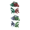
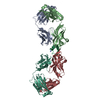
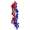
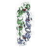
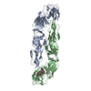
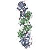
| Title | Complex of the neisserial PorA P1.4 epitope peptide and two Fab- fragments (antibody MN20B9.34) | ||||||
 Components Components |
| ||||||
 Keywords Keywords | IMMUNE SYSTEM / FAB FRAGMENT / ANTI-PORA ANTIBODY / P1.4 ANTIBODY / ANTIGEN / PORIN / TRANSMEMBRANE / ANTIBODY-ANTIGEN COMPLEX | ||||||
| Function / homology |  Function and homology information Function and homology informationporin activity / cell outer membrane / monoatomic ion transmembrane transport Similarity search - Function | ||||||
| Biological species |   NEISSERIA MENINGITIDIS (bacteria) NEISSERIA MENINGITIDIS (bacteria) | ||||||
| Method |  X-RAY DIFFRACTION / SYNCHROTRON / MOLECULAR REPLACEMENT / Resolution: 1.95 Å X-RAY DIFFRACTION / SYNCHROTRON / MOLECULAR REPLACEMENT / Resolution: 1.95 Å | ||||||
 Authors Authors | Oomen, C.J. / Hoogerhout, P. / Kuipers, B. / Vidarsson, G. / Van Alphen, L. / Gros, P. | ||||||
 Citation Citation | Journal: J.Mol.Biol. / Year: 2005 Title: Crystal Structure of an Anti-Meningococcal Subtype P1.4 Pora Antibody Provides Basis for Peptide- Vaccine Design. Authors: Oomen, C.J. / Hoogerhout, P. / Kuipers, B. / Vidarsson, G. / Van Alphen, L. / Gros, P. | ||||||
| History |
| ||||||
| Remark 700 | SHEET THE SHEET STRUCTURE OF THIS MOLECULE IS BIFURCATED. IN ORDER TO REPRESENT THIS FEATURE IN ... SHEET THE SHEET STRUCTURE OF THIS MOLECULE IS BIFURCATED. IN ORDER TO REPRESENT THIS FEATURE IN THE SHEET RECORDS BELOW, TWO SHEETS ARE DEFINED. |
- Structure visualization
| Structure viewer | Molecule: MolmilJmol/JSmol |
|---|
- Downloads & links
-Download
| PDBx/mmCIF format | 2brr.cif.gz | 184.4 KB | Display | PDBx/mmCIF format |
|---|---|---|---|---|
| PDB format | pdb2brr.ent.gz | 146.7 KB | Display | PDB format |
| PDBx/mmJSON format | 2brr.json.gz | Tree view | PDBx/mmJSON format | |
| Others |  Other downloads Other downloads |
-Validation report
| Arichive directory | https://data.pdbj.org/pub/pdb/validation_reports/br/2brrftp://data.pdbj.org/pub/pdb/validation_reports/br/2brr | HTTPS FTP |
|---|
-Related structure data
| Related structure data |  1ce1S S: Starting model for refinement |
|---|---|
| Similar structure data |
-Links
 PDBj
PDBj


- Assembly
| Deposited unit | 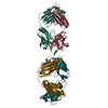
| ||||||||||||||||||||||||||||||||||||||||||||||||||||||||||||||||||||||||||||||||||||||
|---|---|---|---|---|---|---|---|---|---|---|---|---|---|---|---|---|---|---|---|---|---|---|---|---|---|---|---|---|---|---|---|---|---|---|---|---|---|---|---|---|---|---|---|---|---|---|---|---|---|---|---|---|---|---|---|---|---|---|---|---|---|---|---|---|---|---|---|---|---|---|---|---|---|---|---|---|---|---|---|---|---|---|---|---|---|---|---|
| 1 |
| ||||||||||||||||||||||||||||||||||||||||||||||||||||||||||||||||||||||||||||||||||||||
| Unit cell |
| ||||||||||||||||||||||||||||||||||||||||||||||||||||||||||||||||||||||||||||||||||||||
| Noncrystallographic symmetry (NCS) | NCS domain:
NCS domain segments:
NCS ensembles :
| ||||||||||||||||||||||||||||||||||||||||||||||||||||||||||||||||||||||||||||||||||||||
| Details | THE PENTAMERIC ASSEMBLY DESCRIBED HERE IS COMPOSED OF TWO HEAVY/LIGHT CHAIN DIMERS, BOTH BOUND TO THE SINGLE CHAIN P.CHAINS H AND L ARE MARGINALLY MORE TIGHTLY BOUND TO THE PEPTIDE THAN CHAINS X AND Y |
-Components
-Protein/peptide , 1 types, 1 molecules P
| #3: Protein/peptide | Mass: 1244.445 Da / Num. of mol.: 1 Fragment: EXTRACELLULAR LOOP 4 OF PORA SUBTYPE P1.4, RESIDUES 16-26 Source method: obtained synthetically / Details: N-TERMINUS ACETYLATED, C-TERMINUS AMIDATED / Source: (synth.) NEISSERIA MENINGITIDIS (bacteria) / References: UniProt: Q51183 |
|---|
-Antibody , 2 types, 4 molecules HYLX
| #1: Antibody | Mass: 24353.113 Da / Num. of mol.: 2 Source method: isolated from a genetically manipulated source Source: (gene. exp.) #2: Antibody | Mass: 23427.684 Da / Num. of mol.: 2 Source method: isolated from a genetically manipulated source Source: (gene. exp.) |
|---|
-Non-polymers , 3 types, 290 molecules 


| #4: Chemical |  Mass: 96.063 Da / Num. of mol.: 2 / Source method: obtained synthetically / Formula: SO4 Mass: 96.063 Da / Num. of mol.: 2 / Source method: obtained synthetically / Formula: SO4#5: Chemical | ChemComp-ACY / |  Mass: 60.052 Da / Num. of mol.: 1 / Source method: obtained synthetically / Formula: C2H4O2 Mass: 60.052 Da / Num. of mol.: 1 / Source method: obtained synthetically / Formula: C2H4O2#6: Water | ChemComp-HOH / | Mass: 18.015 Da / Num. of mol.: 287 / Source method: isolated from a natural source / Formula: H2O |
|---|
-Details
| Has protein modification | Y |
|---|---|
| Sequence details | THE SEQUENCE OF CHAIN P IS GIVEN BY GENBANK ENTRY CAA78633 |
-Experimental details
-Experiment
| Experiment | Method: X-RAY DIFFRACTION / Number of used crystals: 1 |
|---|
- Sample preparation
| Crystal | Density Matthews: 2.2 Å3/Da / Density % sol: 43 % |
|---|---|
| Crystal grow | pH: 4 Details: 0.2 M AMMONIUM SULPHATE, 0.1 M SODIUM ACETATE/ACETIC ACID (PH 4.0) AND 25% W/V PEG 3000 |
-Data collection
| Diffraction | Mean temperature: 100 K |
|---|---|
| Diffraction source | Source: SYNCHROTRON / Site: ESRF  / Beamline: ID14-3 / Wavelength: 0.9315 / Beamline: ID14-3 / Wavelength: 0.9315 |
| Detector | Type: MARRESEARCH / Detector: CCD / Date: May 10, 2000 |
| Radiation | Protocol: SINGLE WAVELENGTH / Monochromatic (M) / Laue (L): M / Scattering type: x-ray |
| Radiation wavelength | Wavelength: 0.9315 Å / Relative weight: 1 |
| Reflection | Resolution: 1.95→40 Å / Num. obs: 60806 / % possible obs: 96 % / Observed criterion σ(I): 1 / Redundancy: 3.5 % / Rmerge(I) obs: 0.08 / Net I/σ(I): 14.3 |
| Reflection shell | Resolution: 1.95→2.02 Å / Redundancy: 3.4 % / Rmerge(I) obs: 0.39 / Mean I/σ(I) obs: 3.1 / % possible all: 92.1 |
- Processing
| Software |
| ||||||||||||||||||||||||||||||||||||||||||||||||||||||||||||||||||||||||||||||||||||||||||||||||||||||||||||||||||||||||||||||||||||||||||||||||||||||||||||||||||||||||||||||||||||||
|---|---|---|---|---|---|---|---|---|---|---|---|---|---|---|---|---|---|---|---|---|---|---|---|---|---|---|---|---|---|---|---|---|---|---|---|---|---|---|---|---|---|---|---|---|---|---|---|---|---|---|---|---|---|---|---|---|---|---|---|---|---|---|---|---|---|---|---|---|---|---|---|---|---|---|---|---|---|---|---|---|---|---|---|---|---|---|---|---|---|---|---|---|---|---|---|---|---|---|---|---|---|---|---|---|---|---|---|---|---|---|---|---|---|---|---|---|---|---|---|---|---|---|---|---|---|---|---|---|---|---|---|---|---|---|---|---|---|---|---|---|---|---|---|---|---|---|---|---|---|---|---|---|---|---|---|---|---|---|---|---|---|---|---|---|---|---|---|---|---|---|---|---|---|---|---|---|---|---|---|---|---|---|---|
| Refinement | Method to determine structure: MOLECULAR REPLACEMENT Starting model: PDB ENTRY 1CE1 Resolution: 1.95→30 Å / Cor.coef. Fo:Fc: 0.946 / Cor.coef. Fo:Fc free: 0.93 / SU B: 8.458 / SU ML: 0.121 / TLS residual ADP flag: LIKELY RESIDUAL / Cross valid method: THROUGHOUT / ESU R: 0.2 / ESU R Free: 0.17 / Stereochemistry target values: MAXIMUM LIKELIHOOD / Details: HYDROGENS HAVE BEEN ADDED IN THE RIDING POSITIONS.
| ||||||||||||||||||||||||||||||||||||||||||||||||||||||||||||||||||||||||||||||||||||||||||||||||||||||||||||||||||||||||||||||||||||||||||||||||||||||||||||||||||||||||||||||||||||||
| Solvent computation | Ion probe radii: 0.8 Å / Shrinkage radii: 0.8 Å / VDW probe radii: 1.2 Å / Solvent model: MASK | ||||||||||||||||||||||||||||||||||||||||||||||||||||||||||||||||||||||||||||||||||||||||||||||||||||||||||||||||||||||||||||||||||||||||||||||||||||||||||||||||||||||||||||||||||||||
| Displacement parameters | Biso mean: 22.19 Å2
| ||||||||||||||||||||||||||||||||||||||||||||||||||||||||||||||||||||||||||||||||||||||||||||||||||||||||||||||||||||||||||||||||||||||||||||||||||||||||||||||||||||||||||||||||||||||
| Refinement step | Cycle: LAST / Resolution: 1.95→30 Å
| ||||||||||||||||||||||||||||||||||||||||||||||||||||||||||||||||||||||||||||||||||||||||||||||||||||||||||||||||||||||||||||||||||||||||||||||||||||||||||||||||||||||||||||||||||||||
| Refine LS restraints |
|