 Movie
Movie Controller


+ Open data

- Basic information
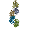
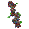
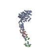
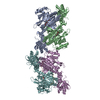
| Entry | Database: PDB / ID: 1afv | ||||||
|---|---|---|---|---|---|---|---|
| Title | HIV-1 CAPSID PROTEIN (P24) COMPLEX WITH FAB25.3 | ||||||
 Components Components |
| ||||||
 Keywords Keywords | Viral protein/Immune system / COMPLEX (VIRAL CAPSID-IMMUNOGLOBULIN) / HIV / CAPSID PROTEIN / P24 / Viral protein-Immune system COMPLEX | ||||||
| Function / homology |  Function and homology information Function and homology informationimmunoglobulin complex / HIV-1 retropepsin / symbiont-mediated activation of host apoptosis / retroviral ribonuclease H / exoribonuclease H / exoribonuclease H activity / DNA integration / viral genome integration into host DNA / establishment of integrated proviral latency / RNA-directed DNA polymerase ...immunoglobulin complex / HIV-1 retropepsin / symbiont-mediated activation of host apoptosis / retroviral ribonuclease H / exoribonuclease H / exoribonuclease H activity / DNA integration / viral genome integration into host DNA / establishment of integrated proviral latency / RNA-directed DNA polymerase / RNA stem-loop binding / viral penetration into host nucleus / host multivesicular body / RNA-directed DNA polymerase activity / RNA-DNA hybrid ribonuclease activity / Transferases; Transferring phosphorus-containing groups; Nucleotidyltransferases / host cell / viral nucleocapsid / DNA recombination / DNA-directed DNA polymerase / aspartic-type endopeptidase activity / Hydrolases; Acting on ester bonds / DNA-directed DNA polymerase activity / adaptive immune response / symbiont-mediated suppression of host gene expression / viral translational frameshifting / symbiont entry into host cell / lipid binding / host cell nucleus / host cell plasma membrane / virion membrane / structural molecule activity / proteolysis / DNA binding / zinc ion binding / metal ion binding Similarity search - Function | ||||||
| Biological species |   Human immunodeficiency virus 1 Human immunodeficiency virus 1 | ||||||
| Method |  X-RAY DIFFRACTION / SYNCHROTRON / MOLECULAR REPLACEMENT, MIR, ANOMALOUS DISPERSION / Resolution: 3.7 Å X-RAY DIFFRACTION / SYNCHROTRON / MOLECULAR REPLACEMENT, MIR, ANOMALOUS DISPERSION / Resolution: 3.7 Å | ||||||
 Authors Authors | Momany, C. / Kovari, L.C. / Prongay, A.J. / Keller, W. / Gitti, R.K. / Lee, B.M. / Gorbalenya, A.E. / Tong, L. / Mcclure, J. / Ehrlich, L.S. ...Momany, C. / Kovari, L.C. / Prongay, A.J. / Keller, W. / Gitti, R.K. / Lee, B.M. / Gorbalenya, A.E. / Tong, L. / Mcclure, J. / Ehrlich, L.S. / Summers, M.F. / Carter, C. / Rossmann, M.G. | ||||||
 Citation Citation | Journal: Nat.Struct.Biol. / Year: 1996 Title: Crystal structure of dimeric HIV-1 capsid protein. Authors: Momany, C. / Kovari, L.C. / Prongay, A.J. / Keller, W. / Gitti, R.K. / Lee, B.M. / Gorbalenya, A.E. / Tong, L. / McClure, J. / Ehrlich, L.S. / Summers, M.F. / Carter, C. / Rossmann, M.G. #1: Journal: Structure / Year: 1995Title: The Use of Antibody Fragments for Crystallization and Structure Determinations Authors: Kovari, L.C. / Momany, C. / Rossmann, M.G. #2: Journal: Proc.Natl.Acad.Sci.USA / Year: 1990Title: Preparation and Crystallization of a Human Immunodeficiency Virus P24-Fab Complex Authors: Prongay, A.J. / Smith, T.J. / Rossmann, M.G. / Ehrlich, L.S. / Carter, C.A. / Mcclure, J. #3: Journal: Aids Res.Hum.Retroviruses / Year: 1990Title: Expression in Escherichia Coli and Purification of Human Immunodeficiency Virus Type 1 Capsid Protein (P24) Authors: Ehrlich, L.S. / Krausslich, H.G. / Wimmer, E. / Carter, C.A. | ||||||
| History |
|
- Structure visualization
| Structure viewer | Molecule: MolmilJmol/JSmol |
|---|
- Downloads & links
-Download
| PDBx/mmCIF format | 1afv.cif.gz | 205.5 KB | Display | PDBx/mmCIF format |
|---|---|---|---|---|
| PDB format | pdb1afv.ent.gz | 160.5 KB | Display | PDB format |
| PDBx/mmJSON format | 1afv.json.gz | Tree view | PDBx/mmJSON format | |
| Others |  Other downloads Other downloads |
-Validation report
| Arichive directory | https://data.pdbj.org/pub/pdb/validation_reports/af/1afvftp://data.pdbj.org/pub/pdb/validation_reports/af/1afv | HTTPS FTP |
|---|
-Related structure data
| Similar structure data |
|---|


-Links
 PDBj
PDBj


- Assembly
| Deposited unit | 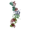
| ||||||||||||||||
|---|---|---|---|---|---|---|---|---|---|---|---|---|---|---|---|---|---|
| 1 |
| ||||||||||||||||
| Unit cell |
| ||||||||||||||||
| Noncrystallographic symmetry (NCS) | NCS oper:
|
-Components
| #1: Protein | Mass: 16716.189 Da / Num. of mol.: 2 / Fragment: AMINO-TERMINAL DOMAIN RESIDUES 1 - 151 Source method: isolated from a genetically manipulated source Source: (gene. exp.) Human immunodeficiency virus 1 / Genus: Lentivirus / Strain: C-12 / Description: IIIB/LAI ISOLATE OF HIV-1; / Cell line: BL21 / Gene: GAG / Plasmid: PHIV-FSII / Species (production host): Escherichia coli / Gene (production host): GAG / Production host:  #2: Antibody | Mass: 23741.270 Da / Num. of mol.: 2 Fragment: LIGHT CHAIN RESIDUES 1 - 217, HEAVY CHAIN RESIDUES 1 - 220 Source method: isolated from a natural source / Source: (natural) #3: Antibody | Mass: 23701.412 Da / Num. of mol.: 2 Fragment: LIGHT CHAIN RESIDUES 1 - 217, HEAVY CHAIN RESIDUES 1 - 220 Source method: isolated from a natural source / Source: (natural) #4: Chemical |   Mass: 207.200 Da / Num. of mol.: 2 / Source method: obtained synthetically / Formula: Pb Mass: 207.200 Da / Num. of mol.: 2 / Source method: obtained synthetically / Formula: PbHas protein modification | Y | |
|---|
-Experimental details
-Experiment
| Experiment | Method: X-RAY DIFFRACTION / Number of used crystals: 1 |
|---|
- Sample preparation
| Crystal | Density Matthews: 2.9 Å3/Da / Density % sol: 57 % | ||||||||||||||||||||||||||||||
|---|---|---|---|---|---|---|---|---|---|---|---|---|---|---|---|---|---|---|---|---|---|---|---|---|---|---|---|---|---|---|---|
| Crystal grow | pH: 7 Details: 16% PEG 3350, 50 MM BISTRIS-HCL, PH 7.0, 0.1% BETA-OCTYLGLUCOSIDE, 1 MM NAN3 | ||||||||||||||||||||||||||||||
| Crystal grow | *PLUS Method: vapor diffusion, sitting drop / PH range low: 7.5 / PH range high: 6 | ||||||||||||||||||||||||||||||
| Components of the solutions | *PLUS
|
-Data collection
| Diffraction | Mean temperature: 120 K |
|---|---|
| Diffraction source | Source: SYNCHROTRON / Site: CHESS  / Beamline: A1 / Wavelength: 0.909 / Beamline: A1 / Wavelength: 0.909 |
| Detector | Type: PRINCETON 2K / Detector: CCD / Date: Dec 1, 1995 |
| Radiation | Monochromatic (M) / Laue (L): M / Scattering type: x-ray |
| Radiation wavelength | Wavelength: 0.909 Å / Relative weight: 1 |
| Reflection | Resolution: 3→100 Å / Num. obs: 29703 / % possible obs: 87.1 % / Observed criterion σ(I): 2 / Redundancy: 3.5 % / Rmerge(I) obs: 0.064 |
| Reflection shell | Resolution: 3→3.11 Å / Rmerge(I) obs: 0.37 / % possible all: 50.4 |
| Reflection shell | *PLUS % possible obs: 50.4 % |
- Processing
| Software |
| ||||||||||||||||||||||||||||||||||||||||||||||||||||||||||||
|---|---|---|---|---|---|---|---|---|---|---|---|---|---|---|---|---|---|---|---|---|---|---|---|---|---|---|---|---|---|---|---|---|---|---|---|---|---|---|---|---|---|---|---|---|---|---|---|---|---|---|---|---|---|---|---|---|---|---|---|---|---|
| Refinement | Method to determine structure: MOLECULAR REPLACEMENT, MIR, ANOMALOUS DISPERSION Starting model: FAB Resolution: 3.7→12 Å / Cross valid method: THROUGHOUT / σ(F): 2 Details: THE CARBOXY-TERMINAL RESIDUES 152 - 231 OF THE HIV-1 CAPSID PROTEIN ARE DISORDERED AND NOT VISIBLE IN THE ELECTRON DENSITY MAPS.
| ||||||||||||||||||||||||||||||||||||||||||||||||||||||||||||
| Displacement parameters | Biso mean: 36.7 Å2 | ||||||||||||||||||||||||||||||||||||||||||||||||||||||||||||
| Refinement step | Cycle: LAST / Resolution: 3.7→12 Å
| ||||||||||||||||||||||||||||||||||||||||||||||||||||||||||||
| Refine LS restraints |
| ||||||||||||||||||||||||||||||||||||||||||||||||||||||||||||
| Refine LS restraints NCS | NCS model details: RESTRAINTS | ||||||||||||||||||||||||||||||||||||||||||||||||||||||||||||
| Software | *PLUS Name: X-PLOR / Version: 3.8 / Classification: refinement | ||||||||||||||||||||||||||||||||||||||||||||||||||||||||||||
| Refinement | *PLUS | ||||||||||||||||||||||||||||||||||||||||||||||||||||||||||||
| Solvent computation | *PLUS | ||||||||||||||||||||||||||||||||||||||||||||||||||||||||||||
| Displacement parameters | *PLUS | ||||||||||||||||||||||||||||||||||||||||||||||||||||||||||||
| Refine LS restraints | *PLUS
|
