 Movie
Movie Controller

[English] 日本語
 Yorodumi
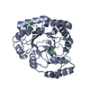
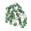
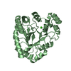
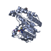
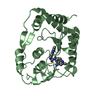
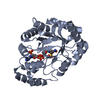
- PDB-1a80: Native 2,5-DIKETO-D-GLUCONIC acid reductase a from CORYNBACTERIUM... -
+ Open data

- Basic information
| Entry | Database: PDB / ID: 1a80 | ||||||
|---|---|---|---|---|---|---|---|
| Title | Native 2,5-DIKETO-D-GLUCONIC acid reductase a from CORYNBACTERIUM SP. complexed with nadph | ||||||
 Components Components | 2,5-DIKETO-D-GLUCONIC ACID REDUCTASE A | ||||||
 Keywords Keywords | OXIDOREDUCTASE / ALPHA8/BETA8 BARREL / 2 / 5-DIKETO-D-GLUCONIC ACID / COMMERCIAL VITAMIN C SYNTHESIS | ||||||
| Function / homology |  Function and homology information Function and homology information2,5-didehydrogluconate reductase (2-dehydro-L-gulonate-forming) / L-ascorbic acid biosynthetic process / oxidoreductase activity, acting on the CH-OH group of donors, NAD or NADP as acceptor / cytoplasm Similarity search - Function | ||||||
| Biological species |  Corynebacterium sp. (bacteria) Corynebacterium sp. (bacteria) | ||||||
| Method |  X-RAY DIFFRACTION / MOLECULAR REPLACEMENT / Resolution: 2.1 Å X-RAY DIFFRACTION / MOLECULAR REPLACEMENT / Resolution: 2.1 Å | ||||||
 Authors Authors | Khurana, S. / Powers, D.B. / Anderson, S. / Blaber, M. | ||||||
 Citation Citation | Journal: Proc.Natl.Acad.Sci.USA / Year: 1998 Title: Crystal structure of 2,5-diketo-D-gluconic acid reductase A complexed with NADPH at 2.1-A resolution. Authors: Khurana, S. / Powers, D.B. / Anderson, S. / Blaber, M. #1: Journal: Biochem.J. / Year: 1997Title: Comparative Anatomy of the Aldo-Keto Reductase Superfamily Authors: Jez, J.M. / Bennett, M.J. / Schlegel, B.P. / Lewis, M. / Penning, T.M. #2: Journal: Science / Year: 1985Title: Production of 2-Keto-L-Gulonate, an Intermediate in L-Ascorbate Synthesis, by a Genetically Modified Erwinia Herbicola Authors: Anderson, S. / Marks, C.B. / Lazarus, R. / Miller, J. / Stafford, K. / Seymour, J. / Light, D. / Rastetter, W. | ||||||
| History |
|
- Structure visualization
| Structure viewer | Molecule: MolmilJmol/JSmol |
|---|
- Downloads & links
-Download
| PDBx/mmCIF format | 1a80.cif.gz | 69.7 KB | Display | PDBx/mmCIF format |
|---|---|---|---|---|
| PDB format | pdb1a80.ent.gz | 50.4 KB | Display | PDB format |
| PDBx/mmJSON format | 1a80.json.gz | Tree view | PDBx/mmJSON format | |
| Others |  Other downloads Other downloads |
-Validation report
| Arichive directory | https://data.pdbj.org/pub/pdb/validation_reports/a8/1a80ftp://data.pdbj.org/pub/pdb/validation_reports/a8/1a80 | HTTPS FTP |
|---|
-Related structure data
| Related structure data | 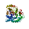 1adsS S: Starting model for refinement |
|---|---|
| Similar structure data |


-Links
 PDBj
PDBj


- Assembly
| Deposited unit | 
| ||||||||
|---|---|---|---|---|---|---|---|---|---|
| 1 |
| ||||||||
| Unit cell |
|
-Components
| #1: Protein | Mass: 30020.473 Da / Num. of mol.: 1 Source method: isolated from a genetically manipulated source Source: (gene. exp.) Corynebacterium sp. (bacteria) / Gene: 2 5-DIKETO-D-GLUCONIC ACID / Variant: A / Plasmid: PTRP1-35 / Gene (production host): 2 5-DIKETO-D-GLUCONIC ACID / Production host: Pantoea citrea (bacteria) / Strain (production host): IFO3263 / References: UniProt: P06632 |
|---|---|
| #2: Chemical | ChemComp-NDP /   Mass: 745.421 Da / Num. of mol.: 1 / Source method: obtained synthetically / Formula: C21H30N7O17P3 Mass: 745.421 Da / Num. of mol.: 1 / Source method: obtained synthetically / Formula: C21H30N7O17P3 |
| #3: Water | ChemComp-HOH /  Mass: 18.015 Da / Num. of mol.: 105 / Source method: isolated from a natural source / Formula: H2O Mass: 18.015 Da / Num. of mol.: 105 / Source method: isolated from a natural source / Formula: H2O |
| Has protein modification | N |
-Experimental details
-Experiment
| Experiment | Method: X-RAY DIFFRACTION / Number of used crystals: 1 |
|---|
- Sample preparation
| Crystal | Density Matthews: 2.16 Å3/Da / Density % sol: 50.31 % | |||||||||||||||||||||||||
|---|---|---|---|---|---|---|---|---|---|---|---|---|---|---|---|---|---|---|---|---|---|---|---|---|---|---|
| Crystal grow | pH: 7.1 / Details: pH 7.1 | |||||||||||||||||||||||||
| Crystal grow | *PLUS Method: vapor diffusion, hanging drop / Details: and 4 degrees centigrade | |||||||||||||||||||||||||
| Components of the solutions | *PLUS
|
-Data collection
| Diffraction | Mean temperature: 295 K |
|---|---|
| Diffraction source | Source: ROTATING ANODE / Type: RIGAKU RUH2R / Wavelength: 1.5418 |
| Detector | Type: RIGAKU RAXIS II / Detector: IMAGE PLATE / Date: Jul 7, 1996 |
| Radiation | Monochromator: GRAPHITE(002) / Monochromatic (M) / Laue (L): M / Scattering type: x-ray |
| Radiation wavelength | Wavelength: 1.5418 Å / Relative weight: 1 |
| Reflection | Resolution: 2.1→100 Å / Num. obs: 14803 / % possible obs: 85 % / Observed criterion σ(I): 0 / Redundancy: 2.6 % / Biso Wilson estimate: 14.6 Å2 / Rsym value: 0.116 / Net I/σ(I): 10 |
| Reflection shell | Resolution: 2.1→2.18 Å / Redundancy: 2.6 % / Mean I/σ(I) obs: 2.5 / Rsym value: 0.421 / % possible all: 75.2 |
| Reflection | *PLUS Rmerge(I) obs: 0.116 |
| Reflection shell | *PLUS % possible obs: 75.2 % / Rmerge(I) obs: 0.421 |
- Processing
| Software |
| ||||||||||||||||||||||||||||||
|---|---|---|---|---|---|---|---|---|---|---|---|---|---|---|---|---|---|---|---|---|---|---|---|---|---|---|---|---|---|---|---|
| Refinement | Method to determine structure: MOLECULAR REPLACEMENT Starting model: PDB ENTRY 1ADS Resolution: 2.1→60 Å / Isotropic thermal model: TRONRUD, 1996 / σ(F): 0 / Stereochemistry target values: TRONRUD, 1987 /
| ||||||||||||||||||||||||||||||
| Solvent computation | Solvent model: TRONRUD, 1987 / Bsol: 365.4 Å2 / ksol: 0.972 e/Å3 | ||||||||||||||||||||||||||||||
| Refinement step | Cycle: LAST / Resolution: 2.1→60 Å
| ||||||||||||||||||||||||||||||
| Refine LS restraints |
| ||||||||||||||||||||||||||||||
| Software | *PLUS Name: TNT / Version: 5E / Classification: refinement | ||||||||||||||||||||||||||||||
| Refinement | *PLUS Rfactor obs: 0.191 / Rfactor Rfree: 0.288 | ||||||||||||||||||||||||||||||
| Solvent computation | *PLUS | ||||||||||||||||||||||||||||||
| Displacement parameters | *PLUS |
