 Movie
Movie Controller

[English] 日本語
 Yorodumi
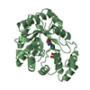
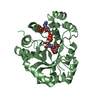
- PDB-1m9h: Corynebacterium 2,5-DKGR A and Phe 22 replaced with Tyr (F22Y), L... -
+ Open data

- Basic information
| Entry | Database: PDB / ID: 1m9h | ||||||
|---|---|---|---|---|---|---|---|
| Title | Corynebacterium 2,5-DKGR A and Phe 22 replaced with Tyr (F22Y), Lys 232 replaced with Gly (K232G), Arg 238 replaced with His (R238H)and Ala 272 replaced with Gly (A272G)in presence of NADH cofactor | ||||||
 Components Components | 2,5-diketo-D-gluconic acid reductase A | ||||||
 Keywords Keywords | OXIDOREDUCTASE / TIM-barrel | ||||||
| Function / homology |  Function and homology information Function and homology information2,5-didehydrogluconate reductase (2-dehydro-L-gulonate-forming) / L-ascorbic acid biosynthetic process / oxidoreductase activity, acting on the CH-OH group of donors, NAD or NADP as acceptor / cytoplasm Similarity search - Function | ||||||
| Biological species |  Corynebacterium sp. (bacteria) Corynebacterium sp. (bacteria) | ||||||
| Method |  X-RAY DIFFRACTION / MOLECULAR REPLACEMENT / Resolution: 2 Å X-RAY DIFFRACTION / MOLECULAR REPLACEMENT / Resolution: 2 Å | ||||||
 Authors Authors | Sanli, G. / Blaber, M. | ||||||
 Citation Citation | Journal: Protein Sci. / Year: 2004 Title: Structural alteration of cofactor specificity in Corynebacterium 2,5-diketo-D-gluconic acid reductase Authors: Sanli, G. / Banta, S. / Anderson, S. / Blaber, M. | ||||||
| History |
|
- Structure visualization
| Structure viewer | Molecule: MolmilJmol/JSmol |
|---|
- Downloads & links
-Download
| PDBx/mmCIF format | 1m9h.cif.gz | 69.4 KB | Display | PDBx/mmCIF format |
|---|---|---|---|---|
| PDB format | pdb1m9h.ent.gz | 50.6 KB | Display | PDB format |
| PDBx/mmJSON format | 1m9h.json.gz | Tree view | PDBx/mmJSON format | |
| Others |  Other downloads Other downloads |
-Validation report
| Arichive directory | https://data.pdbj.org/pub/pdb/validation_reports/m9/1m9hftp://data.pdbj.org/pub/pdb/validation_reports/m9/1m9h | HTTPS FTP |
|---|
-Related structure data
| Related structure data |  1a80S S: Starting model for refinement |
|---|---|
| Similar structure data |


-Links
 PDBj
PDBj
- Assembly
| Deposited unit | 
| ||||||||
|---|---|---|---|---|---|---|---|---|---|
| 1 |
| ||||||||
| 2 | 
| ||||||||
| Unit cell |
|
-Components
| #1: Protein | Mass: 30062.465 Da / Num. of mol.: 1 / Mutation: F22Y, K232G, R238H, A272G Source method: isolated from a genetically manipulated source Source: (gene. exp.) Corynebacterium sp. (bacteria) / Plasmid: pET-21a(+) / Production host: References: UniProt: P06632, Oxidoreductases; Acting on the CH-OH group of donors; With NAD+ or NADP+ as acceptor |
|---|---|
| #2: Chemical | ChemComp-SO4 /   Mass: 96.063 Da / Num. of mol.: 1 / Source method: obtained synthetically / Formula: SO4 Mass: 96.063 Da / Num. of mol.: 1 / Source method: obtained synthetically / Formula: SO4 |
| #3: Chemical | ChemComp-NAD /   Mass: 663.425 Da / Num. of mol.: 1 / Source method: obtained synthetically / Formula: C21H27N7O14P2 / Comment: NAD*YM Mass: 663.425 Da / Num. of mol.: 1 / Source method: obtained synthetically / Formula: C21H27N7O14P2 / Comment: NAD*YM |
| #4: Water | ChemComp-HOH /  Mass: 18.015 Da / Num. of mol.: 146 / Source method: isolated from a natural source / Formula: H2O Mass: 18.015 Da / Num. of mol.: 146 / Source method: isolated from a natural source / Formula: H2O |
-Experimental details
-Experiment
| Experiment | Method: X-RAY DIFFRACTION / Number of used crystals: 2 |
|---|
- Sample preparation
| Crystal | Density Matthews: 2.46 Å3/Da / Density % sol: 50.07 % | ||||||||||||||||||||||||||||||
|---|---|---|---|---|---|---|---|---|---|---|---|---|---|---|---|---|---|---|---|---|---|---|---|---|---|---|---|---|---|---|---|
| Crystal grow | Temperature: 298 K / Method: vapor diffusion, hanging drop / pH: 7.5 Details: lithium sulfate monohydrate, hepes-sodium, pH 7.5, VAPOR DIFFUSION, HANGING DROP, temperature 298K | ||||||||||||||||||||||||||||||
| Crystal grow | *PLUS Method: vapor diffusion, hanging drop | ||||||||||||||||||||||||||||||
| Components of the solutions | *PLUS
|
-Data collection
| Diffraction |
| ||||||||||||||||||
|---|---|---|---|---|---|---|---|---|---|---|---|---|---|---|---|---|---|---|---|
| Diffraction source |
| ||||||||||||||||||
| Detector |
| ||||||||||||||||||
| Radiation |
| ||||||||||||||||||
| Radiation wavelength | Wavelength: 1.5418 Å / Relative weight: 1 | ||||||||||||||||||
| Reflection | Resolution: 2→40 Å / Num. all: 20178 / Num. obs: 17803 / % possible obs: 88.2 % / Observed criterion σ(F): 3 / Observed criterion σ(I): -3 / Redundancy: 4.5 % / Biso Wilson estimate: 15 Å2 / Rmerge(I) obs: 0.114 / Net I/σ(I): 16.6 | ||||||||||||||||||
| Reflection shell | Resolution: 2→2.05 Å / Rmerge(I) obs: 0.276 / Mean I/σ(I) obs: 4.2 / % possible all: 83.3 | ||||||||||||||||||
| Reflection | *PLUS Lowest resolution: 27 Å / Num. obs: 18364 / % possible obs: 92.9 % / Num. measured all: 242073 | ||||||||||||||||||
| Reflection shell | *PLUS % possible obs: 83.3 % |
- Processing
| Software |
| ||||||||||||||||||||
|---|---|---|---|---|---|---|---|---|---|---|---|---|---|---|---|---|---|---|---|---|---|
| Refinement | Method to determine structure: MOLECULAR REPLACEMENT Starting model: PDB ENTRY 1A80 Resolution: 2→40 Å / Cross valid method: THROUGHOUT / σ(F): 3 / σ(I): 3 / Stereochemistry target values: TRONRUD
| ||||||||||||||||||||
| Refinement step | Cycle: LAST / Resolution: 2→40 Å
| ||||||||||||||||||||
| Refine LS restraints |
| ||||||||||||||||||||
| LS refinement shell | Resolution: 2→2.05 Å /
| ||||||||||||||||||||
| Refinement | *PLUS Lowest resolution: 27 Å / Rfactor Rwork: 0.214 | ||||||||||||||||||||
| Solvent computation | *PLUS | ||||||||||||||||||||
| Displacement parameters | *PLUS | ||||||||||||||||||||
| Refine LS restraints | *PLUS
|
