 Movie
Movie Controller


+ Open data

- Basic information
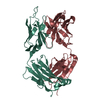
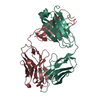
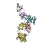
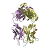
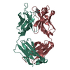
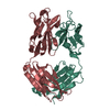
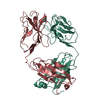
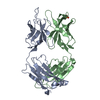
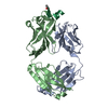
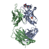
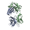
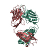
| Entry | Database: PDB / ID: 1ehl | ||||||
|---|---|---|---|---|---|---|---|
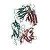
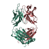
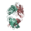
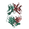
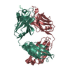
| Title | 64M-2 ANTIBODY FAB COMPLEXED WITH D(5HT)(6-4)T | ||||||
 Components Components |
| ||||||
 Keywords Keywords | IMMUNE SYSTEM / IMMUNOGLOBULIN / PROTEIN-DNA COMPLEX / DNA PHOTOPRODUCT | ||||||
| Function / homology |  Function and homology information Function and homology informationFc-gamma receptor I complex binding / alpha-beta T cell receptor complex / immunoglobulin complex, circulating / immunoglobulin receptor binding / IgG immunoglobulin complex / complement activation, classical pathway / antigen binding / B cell differentiation / antibacterial humoral response / adaptive immune response ...Fc-gamma receptor I complex binding / alpha-beta T cell receptor complex / immunoglobulin complex, circulating / immunoglobulin receptor binding / IgG immunoglobulin complex / complement activation, classical pathway / antigen binding / B cell differentiation / antibacterial humoral response / adaptive immune response / : / extracellular region / plasma membrane Similarity search - Function | ||||||
| Biological species |  | ||||||
| Method |  X-RAY DIFFRACTION / Resolution: 2.4 Å X-RAY DIFFRACTION / Resolution: 2.4 Å | ||||||
 Authors Authors | Yokoyama, H. / Mizutani, R. / Satow, Y. / Komatsu, Y. / Ohtsuka, E. / Nikaido, O. | ||||||
 Citation Citation | Journal: J.Mol.Biol. / Year: 2000 Title: Crystal structure of the 64M-2 antibody Fab fragment in complex with a DNA dT(6-4)T photoproduct formed by ultraviolet radiation. Authors: Yokoyama, H. / Mizutani, R. / Satow, Y. / Komatsu, Y. / Ohtsuka, E. / Nikaido, O. #1: Journal: Photochem.Photobiol. / Year: 1991Title: Simultaneous Establishment of Monoclonal Antibodies Specific for Either Cyclobutane Pyrimidine Dimer or (6-4)photoproduct from the Same Mouse Immunized with Ultraviolet-Irradiated DNA Authors: Mori, T. / Nakane, M. / Hattori, T. / Matsunaga, T. / Ihara, M. / Nikaido, O. #2: Journal: BIOCHIM.BIOPHYS.ACTA / Year: 1998Title: Antibodies Specific for (6-4) DNA Photoproducts: Cloning, Antibody Modeling and Construction of a Single-Chain Fv Derivative Authors: Morioka, H. / Miura, H. / Kobayashi, H. / Koizumi, T. / Fujii, K. / Asano, K. / Matsunaga, T. / Nikaido, O. / Stewart, J.D. / Ohtsuka, E. | ||||||
| History |
|
- Structure visualization
| Structure viewer | Molecule: MolmilJmol/JSmol |
|---|
- Downloads & links
-Download
| PDBx/mmCIF format | 1ehl.cif.gz | 107.2 KB | Display | PDBx/mmCIF format |
|---|---|---|---|---|
| PDB format | pdb1ehl.ent.gz | 80.2 KB | Display | PDB format |
| PDBx/mmJSON format | 1ehl.json.gz | Tree view | PDBx/mmJSON format | |
| Others |  Other downloads Other downloads |
-Validation report
| Arichive directory | https://data.pdbj.org/pub/pdb/validation_reports/eh/1ehlftp://data.pdbj.org/pub/pdb/validation_reports/eh/1ehl | HTTPS FTP |
|---|
-Related structure data
| Similar structure data |
|---|


-Links
 PDBj
PDBj


- Assembly
| Deposited unit | 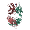
| ||||||||
|---|---|---|---|---|---|---|---|---|---|
| 1 |
| ||||||||
| Unit cell |
|
-Components
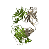
| #1: DNA chain | Mass: 501.464 Da / Num. of mol.: 1 / Source method: obtained synthetically / Details: Purified from ultraviolet-radiated dTpT |
|---|---|
| #2: Antibody | Mass: 23891.496 Da / Num. of mol.: 1 / Source method: isolated from a natural source / Source: (natural) |
| #3: Antibody | Mass: 23522.301 Da / Num. of mol.: 1 / Source method: isolated from a natural source / Source: (natural) |
| #4: Water | ChemComp-HOH /  Mass: 18.015 Da / Num. of mol.: 360 / Source method: isolated from a natural source / Formula: H2O Mass: 18.015 Da / Num. of mol.: 360 / Source method: isolated from a natural source / Formula: H2O |
| Has protein modification | Y |
-Experimental details
-Experiment
| Experiment | Method: X-RAY DIFFRACTION / Number of used crystals: 3 |
|---|
- Sample preparation
| Crystal | Density Matthews: 2.73 Å3/Da / Density % sol: 54.95 % | ||||||||||||||||||||||||||||||||||||||||||
|---|---|---|---|---|---|---|---|---|---|---|---|---|---|---|---|---|---|---|---|---|---|---|---|---|---|---|---|---|---|---|---|---|---|---|---|---|---|---|---|---|---|---|---|
| Crystal grow | Temperature: 293 K / Method: vapor diffusion, hanging drop / pH: 7.5 Details: PEG 8000, nickel chloride, HEPES, pH 7.5, VAPOR DIFFUSION, HANGING DROP, temperature 293K | ||||||||||||||||||||||||||||||||||||||||||
| Crystal | *PLUS Density % sol: 58 % | ||||||||||||||||||||||||||||||||||||||||||
| Crystal grow | *PLUS Temperature: 20 ℃ / PH range low: 8.4 / PH range high: 7.5 | ||||||||||||||||||||||||||||||||||||||||||
| Components of the solutions | *PLUS
|
-Data collection
| Diffraction | Mean temperature: 100 K |
|---|---|
| Diffraction source | Source: ROTATING ANODE / Type: RIGAKU / Wavelength: 1.5418 |
| Detector | Type: RIGAKU / Detector: IMAGE PLATE / Date: Jul 26, 1997 |
| Radiation | Protocol: SINGLE WAVELENGTH / Monochromatic (M) / Laue (L): M / Scattering type: x-ray |
| Radiation wavelength | Wavelength: 1.5418 Å / Relative weight: 1 |
| Reflection | Resolution: 2.4→28.7 Å / Num. all: 77929 / Num. obs: 18790 / % possible obs: 93.5 % / Observed criterion σ(I): 1 / Redundancy: 4.1 % / Biso Wilson estimate: 30.8 Å2 / Rmerge(I) obs: 0.084 / Net I/σ(I): 18.5 |
| Reflection shell | Resolution: 2.4→2.49 Å / Redundancy: 2.7 % / Rmerge(I) obs: 0.221 / Num. unique all: 1571 / % possible all: 79.1 |
| Reflection | *PLUS Num. measured all: 77929 |
| Reflection shell | *PLUS % possible obs: 79.1 % / Num. unique obs: 1571 / Num. measured obs: 4271 / Mean I/σ(I) obs: 3.2 |
- Processing
| Software |
| |||||||||||||||||||||||||
|---|---|---|---|---|---|---|---|---|---|---|---|---|---|---|---|---|---|---|---|---|---|---|---|---|---|---|
| Refinement | Resolution: 2.4→10 Å / σ(F): 1 / Stereochemistry target values: Engh & Huber
| |||||||||||||||||||||||||
| Refinement step | Cycle: LAST / Resolution: 2.4→10 Å
| |||||||||||||||||||||||||
| Refine LS restraints |
| |||||||||||||||||||||||||
| Software | *PLUS Name: X-PLOR / Version: 3.843 / Classification: refinement | |||||||||||||||||||||||||
| Refinement | *PLUS Highest resolution: 2.4 Å / Lowest resolution: 10 Å / σ(F): 1 / % reflection Rfree: 10 % | |||||||||||||||||||||||||
| Solvent computation | *PLUS | |||||||||||||||||||||||||
| Displacement parameters | *PLUS | |||||||||||||||||||||||||
| LS refinement shell | *PLUS Highest resolution: 2.4 Å / Lowest resolution: 2.49 Å / Rfactor Rfree: 0.359 / Rfactor obs: 0.296 |
