 Movie
Movie Controller


+ Open data

- Basic information
| Entry | Database: PDB / ID: 6ru3 | ||||||
|---|---|---|---|---|---|---|---|
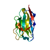
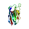
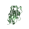
| Title | Crystal structure of the FP specific nanobody hFPNb1 | ||||||
 Components Components | hFP1Nb1 | ||||||
 Keywords Keywords | IMMUNE SYSTEM / antibody / nanobody / single domain antibody | ||||||
| Biological species |  | ||||||
| Method |  X-RAY DIFFRACTION / SYNCHROTRON / MOLECULAR REPLACEMENT / Resolution: 1.26 Å X-RAY DIFFRACTION / SYNCHROTRON / MOLECULAR REPLACEMENT / Resolution: 1.26 Å | ||||||
 Authors Authors | Mazarakis, S.M.M. / Pedersen, D.V. / Laursen, N.S. / Andersen, G.R. | ||||||
| Funding support |  Denmark, 1items Denmark, 1items
| ||||||
 Citation Citation | Journal: Front Immunol / Year: 2019 Title: Structural Basis for Properdin Oligomerization and Convertase Stimulation in the Human Complement System. Authors: Pedersen, D.V. / Gadeberg, T.A.F. / Thomas, C. / Wang, Y. / Joram, N. / Jensen, R.K. / Mazarakis, S.M.M. / Revel, M. / El Sissy, C. / Petersen, S.V. / Lindorff-Larsen, K. / Thiel, S. / ...Authors: Pedersen, D.V. / Gadeberg, T.A.F. / Thomas, C. / Wang, Y. / Joram, N. / Jensen, R.K. / Mazarakis, S.M.M. / Revel, M. / El Sissy, C. / Petersen, S.V. / Lindorff-Larsen, K. / Thiel, S. / Laursen, N.S. / Fremeaux-Bacchi, V. / Andersen, G.R. #1: Journal: Febs Lett. / Year: 2019Title: Crystal structure of peptide-bound neprilysin reveals key binding interactions. Authors: Moss, S. / Subramanian, V. / Acharya, K.R. | ||||||
| History |
|
- Structure visualization
| Structure viewer | Molecule:  MolmilJmol/JSmol MolmilJmol/JSmol |
|---|
- Downloads & links
-Download
| PDBx/mmCIF format | 6ru3.cif.gz | 89.9 KB | Display | PDBx/mmCIF format |
|---|---|---|---|---|
| PDB format | pdb6ru3.ent.gz | 66.1 KB | Display | PDB format |
| PDBx/mmJSON format | 6ru3.json.gz | Tree view | PDBx/mmJSON format | |
| Others |  Other downloads Other downloads |
-Validation report
| Arichive directory | https://data.pdbj.org/pub/pdb/validation_reports/ru/6ru3ftp://data.pdbj.org/pub/pdb/validation_reports/ru/6ru3 | HTTPS FTP |
|---|
-Related structure data
| Related structure data |  6ru5C  6rurC  6rusC  6ruvC  6rv6C  6sejC C: citing same article ( |
|---|---|
| Similar structure data |


-Links
 PDBj
PDBj


- Assembly
| Deposited unit | 
| ||||||||||||
|---|---|---|---|---|---|---|---|---|---|---|---|---|---|
| 1 |
| ||||||||||||
| Unit cell |
| ||||||||||||
| Components on special symmetry positions |
|
-Components
| #1: Antibody | Mass: 14517.155 Da / Num. of mol.: 1 Source method: isolated from a genetically manipulated source Source: (gene. exp.)  |
|---|---|
| #2: Water | ChemComp-HOH /  Mass: 18.015 Da / Num. of mol.: 113 / Source method: isolated from a natural source / Formula: H2O Mass: 18.015 Da / Num. of mol.: 113 / Source method: isolated from a natural source / Formula: H2O |
| Has protein modification | Y |
-Experimental details
-Experiment
| Experiment | Method: X-RAY DIFFRACTION / Number of used crystals: 1 |
|---|
- Sample preparation
| Crystal | Density Matthews: 2.55 Å3/Da / Density % sol: 51.7 % |
|---|---|
| Crystal grow | Temperature: 292 K / Method: vapor diffusion, sitting drop Details: 13.1 % (v/v) MPD, 13.1 % PEG1000 (w/v) 13.1 % (w/v) PEG3350, 100 mM Tris Bicine pH 8.5, 33 mM CaCl2, 33 mM MgCl2, 4 % (v/v) formamide, 114 mM FOS-Choline 8 |
-Data collection
| Diffraction | Mean temperature: 100 K / Serial crystal experiment: N |
|---|---|
| Diffraction source | Source: SYNCHROTRON / Site: PETRA III, EMBL c/o DESY  / Beamline: P13 (MX1) / Wavelength: 0.9763 Å / Beamline: P13 (MX1) / Wavelength: 0.9763 Å |
| Detector | Type: DECTRIS PILATUS 6M / Detector: PIXEL / Date: Nov 1, 2018 |
| Radiation | Protocol: SINGLE WAVELENGTH / Monochromatic (M) / Laue (L): M / Scattering type: x-ray |
| Radiation wavelength | Wavelength: 0.9763 Å / Relative weight: 1 |
| Reflection | Resolution: 1.23037→56.42 Å / Num. obs: 39873 / % possible obs: 98.3 % / Redundancy: 34 % / Biso Wilson estimate: 23 Å2 / Net I/σ(I): 32.3 |
| Reflection shell | Resolution: 1.23037→1.26 Å / Num. unique obs: 2460 / % possible all: 83.4 |
- Processing
| Software |
| |||||||||||||||||||||||||||||||||||||||||||||||||||||||||||||||||||||||||||||||||||||||||||||||||||||||||
|---|---|---|---|---|---|---|---|---|---|---|---|---|---|---|---|---|---|---|---|---|---|---|---|---|---|---|---|---|---|---|---|---|---|---|---|---|---|---|---|---|---|---|---|---|---|---|---|---|---|---|---|---|---|---|---|---|---|---|---|---|---|---|---|---|---|---|---|---|---|---|---|---|---|---|---|---|---|---|---|---|---|---|---|---|---|---|---|---|---|---|---|---|---|---|---|---|---|---|---|---|---|---|---|---|---|---|
| Refinement | Method to determine structure: MOLECULAR REPLACEMENT / Resolution: 1.26→33.85 Å / SU ML: 0.2078 / Cross valid method: FREE R-VALUE / σ(F): 1.33 / Phase error: 27.8807
| |||||||||||||||||||||||||||||||||||||||||||||||||||||||||||||||||||||||||||||||||||||||||||||||||||||||||
| Solvent computation | Shrinkage radii: 0.9 Å / VDW probe radii: 1.11 Å | |||||||||||||||||||||||||||||||||||||||||||||||||||||||||||||||||||||||||||||||||||||||||||||||||||||||||
| Displacement parameters | Biso mean: 31.72 Å2 | |||||||||||||||||||||||||||||||||||||||||||||||||||||||||||||||||||||||||||||||||||||||||||||||||||||||||
| Refinement step | Cycle: LAST / Resolution: 1.26→33.85 Å
| |||||||||||||||||||||||||||||||||||||||||||||||||||||||||||||||||||||||||||||||||||||||||||||||||||||||||
| Refine LS restraints |
| |||||||||||||||||||||||||||||||||||||||||||||||||||||||||||||||||||||||||||||||||||||||||||||||||||||||||
| LS refinement shell |
|
