 Movie
Movie Controller


+ Open data

- Basic information
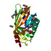
| Entry | Database: PDB / ID: 3t3t | ||||||
|---|---|---|---|---|---|---|---|
| Title | 1.38 A structure of human frataxin variant Q148G | ||||||
 Components Components | Frataxin, mitochondrial | ||||||
 Keywords Keywords | OXIDOREDUCTASE / Fe-S Cluster Biosynthesis / Human mitochondria | ||||||
| Function / homology |  Function and homology information Function and homology informationpositive regulation of lyase activity / proprioception / [4Fe-4S] cluster assembly / Mitochondrial iron-sulfur cluster biogenesis / Complex III assembly / iron chaperone activity / Maturation of TCA enzymes and regulation of TCA cycle / negative regulation of organ growth / mitochondrial respiratory chain complex III assembly / iron-sulfur cluster assembly complex ...positive regulation of lyase activity / proprioception / [4Fe-4S] cluster assembly / Mitochondrial iron-sulfur cluster biogenesis / Complex III assembly / iron chaperone activity / Maturation of TCA enzymes and regulation of TCA cycle / negative regulation of organ growth / mitochondrial respiratory chain complex III assembly / iron-sulfur cluster assembly complex / embryo development ending in birth or egg hatching / Mitochondrial protein import / mitochondrial [2Fe-2S] assembly complex / oxidative phosphorylation / response to iron ion / heme biosynthetic process / adult walking behavior / organ growth / [2Fe-2S] cluster assembly / negative regulation of multicellular organism growth / muscle cell cellular homeostasis / iron-sulfur cluster assembly / negative regulation of release of cytochrome c from mitochondria / ferroxidase / protein autoprocessing / ferroxidase activity / ferric iron binding / iron ion transport / protein maturation / ferrous iron binding / 2 iron, 2 sulfur cluster binding / enzyme activator activity / cellular response to hydrogen peroxide / intracellular iron ion homeostasis / mitochondrial matrix / negative regulation of apoptotic process / mitochondrion / cytosol Similarity search - Function | ||||||
| Biological species |  Homo sapiens (human) Homo sapiens (human) | ||||||
| Method |  X-RAY DIFFRACTION / SYNCHROTRON / MOLECULAR REPLACEMENT / Resolution: 1.38 Å X-RAY DIFFRACTION / SYNCHROTRON / MOLECULAR REPLACEMENT / Resolution: 1.38 Å | ||||||
 Authors Authors | Bridwell-Rabb, J. / Winn, A.M. / Barondeau, D.P. | ||||||
 Citation Citation | Journal: Biochemistry / Year: 2011 Title: Structure-Function Analysis of Friedreich's Ataxia Mutants Reveals Determinants of Frataxin Binding and Activation of the Fe-S Assembly Complex. Authors: Bridwell-Rabb, J. / Winn, A.M. / Barondeau, D.P. | ||||||
| History |
|
- Structure visualization
| Structure viewer | Molecule: MolmilJmol/JSmol |
|---|
- Downloads & links
-Download
| PDBx/mmCIF format | 3t3t.cif.gz | 111.1 KB | Display | PDBx/mmCIF format |
|---|---|---|---|---|
| PDB format | pdb3t3t.ent.gz | 87.2 KB | Display | PDB format |
| PDBx/mmJSON format | 3t3t.json.gz | Tree view | PDBx/mmJSON format | |
| Others |  Other downloads Other downloads |
-Validation report
| Arichive directory | https://data.pdbj.org/pub/pdb/validation_reports/t3/3t3tftp://data.pdbj.org/pub/pdb/validation_reports/t3/3t3t | HTTPS FTP |
|---|
-Related structure data


-Links
 PDBj
PDBj

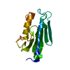
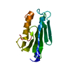
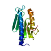
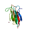
- Assembly
| Deposited unit | 
| ||||||||
|---|---|---|---|---|---|---|---|---|---|
| 1 | 
| ||||||||
| 2 | 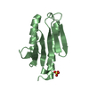
| ||||||||
| 3 | 
| ||||||||
| 4 | 
| ||||||||
| Unit cell |
|
-Components
| #1: Protein | Mass: 14124.570 Da / Num. of mol.: 4 / Fragment: mature form (UNP residues 82-210) / Mutation: Q148G Source method: isolated from a genetically manipulated source Source: (gene. exp.) Homo sapiens (human) / Gene: FXN, FRDA, X25 / Production host:  #2: Chemical | ChemComp-SO4 /   Mass: 96.063 Da / Num. of mol.: 4 / Source method: obtained synthetically / Formula: SO4 Mass: 96.063 Da / Num. of mol.: 4 / Source method: obtained synthetically / Formula: SO4#3: Water | ChemComp-HOH / |  Mass: 18.015 Da / Num. of mol.: 341 / Source method: isolated from a natural source / Formula: H2O Mass: 18.015 Da / Num. of mol.: 341 / Source method: isolated from a natural source / Formula: H2O |
|---|
-Experimental details
-Experiment
| Experiment | Method: X-RAY DIFFRACTION / Number of used crystals: 1 |
|---|
- Sample preparation
| Crystal | Density Matthews: 2.53 Å3/Da / Density % sol: 51.37 % |
|---|---|
| Crystal grow | Temperature: 295 K / Method: vapor diffusion, hanging drop / pH: 5.5 Details: 2.0 M ammonium sulfate, 0.1 M citrate, pH 5.5, VAPOR DIFFUSION, HANGING DROP, temperature 295K |
-Data collection
| Diffraction source | Source: SYNCHROTRON / Site: SSRL  / Beamline: BL11-1 / Wavelength: 1.0971 Å / Beamline: BL11-1 / Wavelength: 1.0971 Å |
|---|---|
| Detector | Type: MARMOSAIC 325 mm CCD / Detector: CCD |
| Radiation | Monochromator: Side scattering bent cube-root I-beam single crystal; asymmetric cut 4.965 degs Protocol: SINGLE WAVELENGTH / Monochromatic (M) / Laue (L): M / Scattering type: x-ray |
| Radiation wavelength | Wavelength: 1.0971 Å / Relative weight: 1 |
| Reflection | Resolution: 1.38→26.19 Å / Num. obs: 111516 / % possible obs: 96.7 % |
| Reflection shell | Resolution: 1.38→1.416 Å / % possible all: 94.24 |
- Processing
| Software |
| |||||||||||||||||||||||||||||||||||||||||||||||||||||||||||||||||
|---|---|---|---|---|---|---|---|---|---|---|---|---|---|---|---|---|---|---|---|---|---|---|---|---|---|---|---|---|---|---|---|---|---|---|---|---|---|---|---|---|---|---|---|---|---|---|---|---|---|---|---|---|---|---|---|---|---|---|---|---|---|---|---|---|---|---|
| Refinement | Method to determine structure: MOLECULAR REPLACEMENT / Resolution: 1.38→26.19 Å / Cor.coef. Fo:Fc: 0.963 / Cor.coef. Fo:Fc free: 0.951 / SU B: 0.979 / SU ML: 0.04 / Cross valid method: THROUGHOUT / ESU R: 0.061 / ESU R Free: 0.064 / Stereochemistry target values: MAXIMUM LIKELIHOOD Details: HYDROGENS HAVE BEEN ADDED IN THE RIDING POSITIONS U VALUES : REFINED INDIVIDUALLY
| |||||||||||||||||||||||||||||||||||||||||||||||||||||||||||||||||
| Solvent computation | Ion probe radii: 0.8 Å / Shrinkage radii: 0.8 Å / VDW probe radii: 1.4 Å / Solvent model: MASK | |||||||||||||||||||||||||||||||||||||||||||||||||||||||||||||||||
| Displacement parameters | Biso mean: 15.332 Å2
| |||||||||||||||||||||||||||||||||||||||||||||||||||||||||||||||||
| Refinement step | Cycle: LAST / Resolution: 1.38→26.19 Å
| |||||||||||||||||||||||||||||||||||||||||||||||||||||||||||||||||
| Refine LS restraints |
| |||||||||||||||||||||||||||||||||||||||||||||||||||||||||||||||||
| LS refinement shell | Resolution: 1.38→1.416 Å / Total num. of bins used: 20
|
