 Movie
Movie Controller

[English] 日本語
 Yorodumi
- PDB-1koa: TWITCHIN KINASE FRAGMENT (C.ELEGANS), AUTOREGULATED PROTEIN KINAS... -
+ Open data

- Basic information
| Entry | Database: PDB / ID: 1koa | ||||||
|---|---|---|---|---|---|---|---|
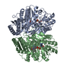
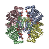
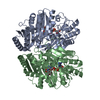
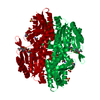
| Title | TWITCHIN KINASE FRAGMENT (C.ELEGANS), AUTOREGULATED PROTEIN KINASE AND IMMUNOGLOBULIN DOMAINS | ||||||
 Components Components | TWITCHIN | ||||||
 Keywords Keywords | KINASE / TWITCHIN / INTRASTERIC REGULATION | ||||||
| Function / homology |  Function and homology information Function and homology informationpositive regulation of sarcomere organization / positive regulation of striated muscle contraction / negative regulation of cell division / positive regulation of locomotion / A band / M band / sarcomere organization / adult locomotory behavior / calmodulin binding / non-specific serine/threonine protein kinase ...positive regulation of sarcomere organization / positive regulation of striated muscle contraction / negative regulation of cell division / positive regulation of locomotion / A band / M band / sarcomere organization / adult locomotory behavior / calmodulin binding / non-specific serine/threonine protein kinase / protein serine kinase activity / protein serine/threonine kinase activity / enzyme binding / ATP binding / metal ion binding Similarity search - Function | ||||||
| Biological species |  | ||||||
| Method |  X-RAY DIFFRACTION / SYNCHROTRON / Resolution: 3.3 Å X-RAY DIFFRACTION / SYNCHROTRON / Resolution: 3.3 Å | ||||||
 Authors Authors | Kobe, B. / Heierhorst, J. / Feil, S.C. / Parker, M.W. / Benian, G.M. / Weiss, K.R. / Kemp, B.E. | ||||||
 Citation Citation | Journal: EMBO J. / Year: 1996 Title: Giant protein kinases: domain interactions and structural basis of autoregulation. Authors: Kobe, B. / Heierhorst, J. / Feil, S.C. / Parker, M.W. / Benian, G.M. / Weiss, K.R. / Kemp, B.E. #1: Journal: Nature / Year: 1996Title: Ca2+/S100 Regulation of Giant Protein Kinases Authors: Heierhorst, J. / Kobe, B. / Feil, S.C. / Parker, M.W. / Benian, G.M. / Weiss, K.R. / Kemp, B.E. | ||||||
| History |
|
- Structure visualization
| Structure viewer | Molecule: MolmilJmol/JSmol |
|---|
- Downloads & links
-Download
| PDBx/mmCIF format | 1koa.cif.gz | 98.6 KB | Display | PDBx/mmCIF format |
|---|---|---|---|---|
| PDB format | pdb1koa.ent.gz | 73.1 KB | Display | PDB format |
| PDBx/mmJSON format | 1koa.json.gz | Tree view | PDBx/mmJSON format | |
| Others |  Other downloads Other downloads |
-Validation report
| Arichive directory | https://data.pdbj.org/pub/pdb/validation_reports/ko/1koaftp://data.pdbj.org/pub/pdb/validation_reports/ko/1koa | HTTPS FTP |
|---|
-Related structure data


-Links
 PDBj
PDBj


- Assembly
| Deposited unit | 
| ||||||||
|---|---|---|---|---|---|---|---|---|---|
| 1 | 
| ||||||||
| Unit cell |
|
-Components
| #1: Protein | Mass: 56426.305 Da / Num. of mol.: 1 / Fragment: KINASE FRAGMENT Source method: isolated from a genetically manipulated source Source: (gene. exp.)  |
|---|
-Experimental details
-Experiment
| Experiment | Method: X-RAY DIFFRACTION |
|---|
- Sample preparation
| Crystal | Density Matthews: 5.52 Å3/Da / Density % sol: 77.71 % | ||||||||||||||||||||||||||||||
|---|---|---|---|---|---|---|---|---|---|---|---|---|---|---|---|---|---|---|---|---|---|---|---|---|---|---|---|---|---|---|---|
| Crystal grow | *PLUS pH: 7.4 / Method: vapor diffusion, hanging drop | ||||||||||||||||||||||||||||||
| Components of the solutions | *PLUS
|
-Data collection
| Diffraction source | Source: SYNCHROTRON / Site: Photon Factory  / Beamline: BL-6A / Wavelength: 1 / Beamline: BL-6A / Wavelength: 1 |
|---|---|
| Detector | Detector: IMAGE PLATE / Date: 1995 |
| Radiation | Monochromatic (M) / Laue (L): M / Scattering type: x-ray |
| Radiation wavelength | Wavelength: 1 Å / Relative weight: 1 |
| Reflection | Num. obs: 17405 / % possible obs: 83.4 % / Observed criterion σ(I): -3 / Redundancy: 4.8 % / Biso Wilson estimate: 100 Å2 / Rmerge(I) obs: 0.157 |
| Reflection | *PLUS Highest resolution: 3.3 Å / Lowest resolution: 3.7 Å / Num. measured all: 39131 |
- Processing
| Software |
| ||||||||||||||||||||||||||||||||||||||||||||||||||||||||||||
|---|---|---|---|---|---|---|---|---|---|---|---|---|---|---|---|---|---|---|---|---|---|---|---|---|---|---|---|---|---|---|---|---|---|---|---|---|---|---|---|---|---|---|---|---|---|---|---|---|---|---|---|---|---|---|---|---|---|---|---|---|---|
| Refinement | Resolution: 3.3→40 Å / σ(F): 0 Details: B-FACTORS WERE REFINED AS 2 GROUPS PER RESIDUE (MAIN-CHAIN ATOMS, SIDE-CHAIN ATOMS).
| ||||||||||||||||||||||||||||||||||||||||||||||||||||||||||||
| Displacement parameters | Biso mean: 88.5 Å2 | ||||||||||||||||||||||||||||||||||||||||||||||||||||||||||||
| Refine analyze |
| ||||||||||||||||||||||||||||||||||||||||||||||||||||||||||||
| Refinement step | Cycle: LAST / Resolution: 3.3→40 Å
| ||||||||||||||||||||||||||||||||||||||||||||||||||||||||||||
| Refine LS restraints |
| ||||||||||||||||||||||||||||||||||||||||||||||||||||||||||||
| Software | *PLUS Name: X-PLOR / Version: 3.1 / Classification: refinement | ||||||||||||||||||||||||||||||||||||||||||||||||||||||||||||
| Refinement | *PLUS Rfactor Rfree: 0.3425 / Rfactor Rwork: 0.2719 | ||||||||||||||||||||||||||||||||||||||||||||||||||||||||||||
| Solvent computation | *PLUS | ||||||||||||||||||||||||||||||||||||||||||||||||||||||||||||
| Displacement parameters | *PLUS |
