 Movie
Movie Controller

[English] 日本語
 Yorodumi
- PDB-1epf: CRYSTAL STRUCTURE OF THE TWO N-TERMINAL IMMUNOGLOBULIN DOMAINS OF... -
+ Open data

- Basic information
| Entry | Database: PDB / ID: 1epf | ||||||
|---|---|---|---|---|---|---|---|
| Title | CRYSTAL STRUCTURE OF THE TWO N-TERMINAL IMMUNOGLOBULIN DOMAINS OF THE NEURAL CELL ADHESION MOLECULE (NCAM) | ||||||
 Components Components | PROTEIN (NEURAL CELL ADHESION MOLECULE) | ||||||
 Keywords Keywords | CELL ADHESION / NCAM / IMMUNOGLOBULIN FOLD / GLYCOPROTEIN | ||||||
| Function / homology |  Function and homology information Function and homology informationNCAM1 interactions / regulation of exocyst assembly / medium-term memory / regulation of semaphorin-plexin signaling pathway / cellular response to fluoride / calcium-independent cell-cell adhesion / peripheral nervous system axon regeneration / homotypic cell-cell adhesion / NCAM signaling for neurite out-growth / Signal transduction by L1 ...NCAM1 interactions / regulation of exocyst assembly / medium-term memory / regulation of semaphorin-plexin signaling pathway / cellular response to fluoride / calcium-independent cell-cell adhesion / peripheral nervous system axon regeneration / homotypic cell-cell adhesion / NCAM signaling for neurite out-growth / Signal transduction by L1 / cellular response to molecule of bacterial origin / fibroblast growth factor receptor binding / thalamus development / commissural neuron axon guidance / behavioral response to ethanol / axonal fasciculation / RAF/MAP kinase cascade / negative regulation of programmed cell death / LRR domain binding / response to morphine / animal organ regeneration / epithelial to mesenchymal transition / cellular response to ethanol / multicellular organismal response to stress / neuron development / phosphatase binding / positive regulation of cardiac muscle cell proliferation / cytoskeletal protein binding / positive regulation of calcium-mediated signaling / response to activity / response to cocaine / response to lead ion / modulation of chemical synaptic transmission / Schaffer collateral - CA1 synapse / neuron projection development / cell-cell junction / heparin binding / growth cone / response to oxidative stress / presynaptic membrane / learning or memory / postsynaptic membrane / cell surface receptor signaling pathway / cell adhesion / neuron projection / response to xenobiotic stimulus / external side of plasma membrane / axon / neuronal cell body / glutamatergic synapse / cell surface / plasma membrane / cytoplasm / cytosol Similarity search - Function | ||||||
| Biological species |  | ||||||
| Method |  X-RAY DIFFRACTION / SYNCHROTRON / Resolution: 1.85 Å X-RAY DIFFRACTION / SYNCHROTRON / Resolution: 1.85 Å | ||||||
 Authors Authors | Kasper, C. / Rasmussen, H. / Kastrup, J.S. / Ikemizu, S. / Jones, E.Y. / Berezin, V. / Bock, E. / Larsen, I.K. | ||||||
 Citation Citation | Journal: Nat.Struct.Biol. / Year: 2000 Title: Structural basis of cell-cell adhesion by NCAM. Authors: Kasper, C. / Rasmussen, H. / Kastrup, J.S. / Ikemizu, S. / Jones, E.Y. / Berezin, V. / Bock, E. / Larsen, I.K. #1: Journal: Acta Crystallogr.,Sect.D / Year: 1999Title: Expression, Crystallization and Preliminary X-Ray Analysis of the Two Amino-Terminal Ig Domains of the Neural Cell Adhesion Molecule (Ncam) Authors: Kasper, C. / Rasmussen, H. / Berezin, V. / Bock, E. / Larsen, I.K. | ||||||
| History |
|
- Structure visualization
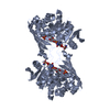
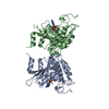
| Structure viewer | Molecule: MolmilJmol/JSmol |
|---|
- Downloads & links
-Download
| PDBx/mmCIF format | 1epf.cif.gz | 197.5 KB | Display | PDBx/mmCIF format |
|---|---|---|---|---|
| PDB format | pdb1epf.ent.gz | 156.6 KB | Display | PDB format |
| PDBx/mmJSON format | 1epf.json.gz | Tree view | PDBx/mmJSON format | |
| Others |  Other downloads Other downloads |
-Validation report
| Arichive directory | https://data.pdbj.org/pub/pdb/validation_reports/ep/1epfftp://data.pdbj.org/pub/pdb/validation_reports/ep/1epf | HTTPS FTP |
|---|
-Related structure data
| Similar structure data |
|---|


-Links
 PDBj
PDBj


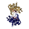
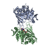
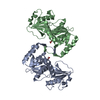
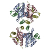
- Assembly
| Deposited unit | 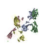
| ||||||||
|---|---|---|---|---|---|---|---|---|---|
| 1 | 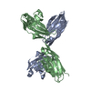
| ||||||||
| 2 | 
| ||||||||
| Unit cell |
|
-Components
| #1: Protein | Mass: 21407.326 Da / Num. of mol.: 4 / Fragment: N-TERMINAL IG DOMAIN Source method: isolated from a genetically manipulated source Source: (gene. exp.)  Pichia pastoris (fungus) / References: UniProt: P13596 Pichia pastoris (fungus) / References: UniProt: P13596#2: Chemical | ChemComp-CA / |   Mass: 40.078 Da / Num. of mol.: 1 / Source method: obtained synthetically / Formula: Ca Mass: 40.078 Da / Num. of mol.: 1 / Source method: obtained synthetically / Formula: Ca#3: Water | ChemComp-HOH / |  Mass: 18.015 Da / Num. of mol.: 1418 / Source method: isolated from a natural source / Formula: H2O Mass: 18.015 Da / Num. of mol.: 1418 / Source method: isolated from a natural source / Formula: H2OHas protein modification | Y | |
|---|
-Experimental details
-Experiment
| Experiment | Method: X-RAY DIFFRACTION / Number of used crystals: 1 |
|---|
- Sample preparation
| Crystal | Density Matthews: 2.43 Å3/Da / Density % sol: 49.33 % | ||||||||||||||||||||||||||||||||||||
|---|---|---|---|---|---|---|---|---|---|---|---|---|---|---|---|---|---|---|---|---|---|---|---|---|---|---|---|---|---|---|---|---|---|---|---|---|---|
| Crystal grow | pH: 6.5 / Details: pH 6.50 | ||||||||||||||||||||||||||||||||||||
| Crystal grow | *PLUS Temperature: 293 K / pH: 7.5 / Method: vapor diffusion, hanging dropDetails: used macroseeding, Kasper, C., (1999) Acta Crystallogr., Sect.D, 55, 1598. | ||||||||||||||||||||||||||||||||||||
| Components of the solutions | *PLUS
|
-Data collection
| Diffraction | Mean temperature: 120 K |
|---|---|
| Diffraction source | Source: SYNCHROTRON / Site: ESRF  / Beamline: BM14 / Wavelength: 0.855 / Beamline: BM14 / Wavelength: 0.855 |
| Detector | Type: MARRESEARCH / Detector: IMAGE PLATE / Date: Aug 28, 1998 |
| Radiation | Protocol: SINGLE WAVELENGTH / Monochromatic (M) / Laue (L): M / Scattering type: x-ray |
| Radiation wavelength | Wavelength: 0.855 Å / Relative weight: 1 |
| Reflection | Resolution: 1.85→30 Å / Num. obs: 64828 / % possible obs: 92.6 % / Observed criterion σ(I): 0 / Redundancy: 2.9 % / Biso Wilson estimate: 18.9 Å2 / Rmerge(I) obs: 0.047 / Net I/σ(I): 21.9 |
| Reflection shell | Resolution: 1.85→1.92 Å / Rmerge(I) obs: 0.308 / % possible all: 90 |
| Reflection | *PLUS Highest resolution: 1.85 Å / Lowest resolution: 30 Å / Observed criterion σ(I): 0 / Redundancy: 2.9 % / Num. measured all: 186351 |
| Reflection shell | *PLUS % possible obs: 90 % / Mean I/σ(I) obs: 3.4 |
- Processing
| Software |
| ||||||||||||||||||||||||||||||||||||||||||||||||||||||||||||
|---|---|---|---|---|---|---|---|---|---|---|---|---|---|---|---|---|---|---|---|---|---|---|---|---|---|---|---|---|---|---|---|---|---|---|---|---|---|---|---|---|---|---|---|---|---|---|---|---|---|---|---|---|---|---|---|---|---|---|---|---|---|
| Refinement | Resolution: 1.85→30 Å / Cross valid method: THROUGHOUT / σ(F): 0 / Stereochemistry target values: ENGH & HUBER
| ||||||||||||||||||||||||||||||||||||||||||||||||||||||||||||
| Refinement step | Cycle: LAST / Resolution: 1.85→30 Å
| ||||||||||||||||||||||||||||||||||||||||||||||||||||||||||||
| Refine LS restraints |
| ||||||||||||||||||||||||||||||||||||||||||||||||||||||||||||
| Software | *PLUS Name: CNS / Classification: refinement | ||||||||||||||||||||||||||||||||||||||||||||||||||||||||||||
| Refinement | *PLUS % reflection Rfree: 5 % | ||||||||||||||||||||||||||||||||||||||||||||||||||||||||||||
| Solvent computation | *PLUS | ||||||||||||||||||||||||||||||||||||||||||||||||||||||||||||
| Displacement parameters | *PLUS | ||||||||||||||||||||||||||||||||||||||||||||||||||||||||||||
| Refine LS restraints | *PLUS
|
